

 PDF
PDF ePub
ePub Citation
 Print
Print


INTRODUCTION
Jarcho-Levin syndrome (JLS) is a rare autosomal-recessive disorder and is a type of segmental costovertebral malformation (1). This syndrome is usually diagnosed in the fetal period by prenatal ultrasonography or in newborns (2). It represents a spectrum of short-trunk skeletal dysplasias with variable involvement of the vertebrae and ribs. Patients with JLS usually die while newborn or in infancy because the small size of their thorax frequently causes respiratory compromise (1, 3, 4). Other abnormalities described in JLS include neural tube defects, Arnold-Chiari malformation and urinary tract abnormalities (2, 5).
The term diastematomyelia refers to a congenital malformation with a longitudinal split in the spinal cord (1). Only six cases of JLS with diastematomyelia have been reported (1, 3). All cases were diagnosed before or at birth, or in childhood. No case involving an adult patient has been reported. The exact life-span of the six reported cases of JLS with diastematomyelia are not known.
We report the seventh case of JLS with diastematomyelia. It is the first case involving an adult patient.
CASE REPORT
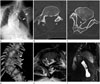
A 37-year-old female visited our hospital for consultation for surgical treatment of thoracolumbar scoliosis. She had a short trunk and neck, with relatively long arms and a slightly protuberant abdomen. Whole spine scanogram showed thoracolumbar scoliosis and extensive malsegmentation of the thoracolumbar spines. Numerical and various intrinsic rib anomalies including irregular fusion and irregular narrowing were also noted on the left side (Fig. 1A).
Axial computed tomography of the spine using an Aquilion 64 (Toshiba Medical Systems, Tokyo, Japan) revealed the spina bifida at the level of the L3 vertebra (Fig. 1B) and diastematomyelia with a large intraspinal bony septum at the L4 vertebral level (Fig. 1C). Volume rendering image reconstruction using an Aquarius iNuition ver 4.4.7 (Terarecon, Foster City, CA, USA) demonstrated a variety of vertebral deformities including hemivertebrae, block vertebrae, butterfly vertebrae and tripedicular vertebra at thoracolumbar spines (Fig. 1D). Abnormal fusion of the left ribs was also noted. The findings were compatible with spondylocostal dysostosis. There was evidence of diastematomyelia with a large intraspinal bony bar at the L4 level. Spinal magnetic resonance images demonstrated the split cord malformation at the whole thoracolumbar vertebral level. Axial T2-weighted scan at the level of T2 showed the separated spinal cord into two hemicords (Fig. 1E). Coronal T2-weighted image revealed a large intraspinal bony septum (Fig. 1F) with separation of the spinal cord into two hemicords.
The patient refused surgery for the thoracolumbar scoliosis.
DISCUSSION
In 1938, Jarcho and Levin (6) first described cases of two siblings with multiple congenital vertebral and rib cage malformations leading to short neck and short thorax. In 1978, Solomon et al. (7) classified cases of JLS into two phenotypic groups based on the presence or absence of rib malformations: spondylothoracic dysostosis (STD) and spondylocostal dysostosis (SCD).
STD is a phenotype of JLS with the absence of rib malformation. It is characterized by segmentation and formation defects in spine, such as hemivertebrae, block vertebrae and unsegmented bars. Patients feature a 'crab-like' chest due to fusion of all the ribs at the costovertebral junction (2). Intrinsic rib malformations have not been reported. SCD has vertebral anomalies and rib malformations that include broadening, bifurcation, abnormal orientation and fusion. Flaring of iliac bones is also present. The incidence of STD is unknown, but the reported prevalence is 0.2 per 100000 liveborns (3, 4).
STD has a poorer prognosis than SCD, because of respiratory complications like restrictive lung disease (2, 3, 4). Dane et al. (2) reported 45% mortality in neonatal or infancy with STD from such respiratory complications. Patients with SCD usually have a much lower mortality rate (3).
Neural tube defects like spina bifida and menigomyelocele are reportedly associated with JLS (2, 8), but cases with diastematomyelia are extremely rare (2). Split cord malformations and related malformations occur as a result of embryological failure of midline axial integration during gastrulation (9).
In 1970, Eller and Morton (10) first reported a case in which diastematomyelia occurred in association with findings of JLS in an infant born to a woman who abused lysergic acid diethylamide during pregnancy. In 2003, Etus et al. (8) reported a case of JLS with diastematomyelia in a 7-year-old girl; she is the oldest patient with JLS and diastematomyelia among the patients reported so far. All reported cases of JLS with diastematomyelia were diagnosed at birth or in childhood. No adult case has hitherto been reported.
We report the seventh case of JLS with diastematomyelia. The patient is the oldest to date. Our case is significant, because the exact lifespans of the reported six cases of JLS with diastematomyelia are not known.
 XML Download