

 PDF
PDF ePub
ePub Citation
 Print
Print


INTRODUCTION
Various kinds of foreign bodies in the pleural space have been reported, and most of them were surgically removed (1). The surface coating polyurethane film of the Terumo wire peeled off during thoracentesis, and its fragments remained in the pyothorax in the form of strings in the reported case. We removed the iatrogenic foreign body in the pyothorax by an interventional technique using a snare catheter and endoscopic biopsy forcep.
CASE REPORT
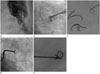
A 62-year-old male was admitted to our hospital with pneumonia and pyothorax. He was referred to our clinic for a diagnostic and therapeutic thoracentesis of the left pyothorax. We attempted percutaneous drainage catheter insertion into the left pyothorax. Under ultrasonography guidance, an 18-gauge aspiration needle (Chiba needle, Cook Medical, Bloomington, IN, USA) was advanced into the pyothorax. Next, under fluoroscopic guidance a 0.035 guide wire (A Plus guide wire, A&A M.D., Gyeonggi, Korea) was passed through the aspiration needle into the pyothorax. However, in the process of wire manipulation, the wire became stuck in the edge of the needle. When the wire was forcedly pulled out, the surface coating polyurethane film of the wire was peeled off, and its fragments remained in the pyothorax in the form of strings. A chest PA and CT scan were performed to determine the exact anatomy and location of the fragments. The chest PA (Fig. 1A) showed linear radiopaque foreign body shadows in the left pyothorax. Surgical removal was not considered for the patient due to diabetes mellitus and hypertension.
Two days later interventional removal was attempted. First, a 10 French 10 cm vascular sheath (Pinnacle introducer sheath, Terumo Medical, Somerset, NJ, USA) was inserted into the left pyothorax, and its tip was positioned near the foreign bodies. Next, a 15 mm diameter snare catheter (Amplatz Goose Neck snare, ev3 Endovascular, Plymouth, MN, USA) was passed through the sheath, and the polyurethane fragments were snatched by the snare catheter and subsequently pulled out through the sheath. The process was repeated several times (Fig. 1B, C). However, we were unable to approach the last remaining fragment with the snare catheter. The sheath was repositioned to find the shortest route to the remaining piece, and removal with the snare catheter was attempted once more. However, there was not enough space for the snare catheter to make a loop around the fragment, resulting in failure to retrieve the final fragment. At that time, we decided to try using the colonoscopic biopsy forcep (Maxum Reusable Forcep, Cook Medical, Bloomington, IN, USA). We inserted the forcep through the sheath and positioned its tip just above the fragment. We then used the forcep to capture the fragment and successfully pull it out through the sheath (Fig. 1D, E). After removal of the foreign bodies, a 10-Fr pigtail catheter was inserted in the left pyothorax. There were no immediate procedure related complications, and no change in hemodynamic or respiratory status during the entire procedure.
DISCUSSION
An empyema is pus in the pleural space. Most empyemas are fibrinopurulent, and characterized by loculated and turbid fluid collections. Advanced empyemas involve organization of the infected pleural effusion. For drainage of an advanced small empyema, we used the Seldinger technique, with an 18-gauge aspiration needle and a 0.035 guide wire (A Plus guide wire, A&A M.D., Gyeonggi, Korea). Using the aspiration needle and a guide wire for the purpose of draining an uncomplicated effusion does not cause any problems; however, in advanced small empyema as in the current case, it can be a big problem. During wire manipulation in such a small, stiff space, the wire can become stuck and scrape on the sharp edge of the needle tip. The surface of the guide wire was coated with polyurethane film, in contrast with the stiff guide wire, hence the surface coating film was easily peeled off. Similar cases can occur in a femoral artery puncture for angiography. If the guide wire is stuck in the Seldinger needle, its surface coating of polyurethane film could likewise be peeled off. Usually, this causes an embolism in the ipsilateral popliteal artery. We have experience removing it using a snare catheter.
Various kinds of foreign bodies in the pleural space have been reported. Most of them have been surgically removed by procedures such as a thoracotomy, video-assisted thoracoscopy, direct pleuroscopy, or simple incision (1, 2, 3, 4, 5). Recently, one case was reported in which a pigtail catheter fragment in the pleural space was removed by an interventional technique using a guide wire (Bentson wire, Cook Medical, Bloomington, IN, USA) and a loop snare (EN Snare Endovascular Snare System, Merit Medical, South Jordan, UT, USA) (6).
In summary, we do not recommend the use of a 0.035 hydrophilic coated guide wire for the purpose of draining advanced small empyema, since it involves a risk of the polyurethane coat peeling off. Pleural foreign bodies are typically removed surgically, but various interventional techniques can also be used as a treatment of choice.
 XML Download