

 PDF
PDF ePub
ePub Citation
 Print
Print


INTRODUCTION
In embryology, two renal veins (ventral and dorsal) are formed by the fusion of the supra- and sub-cardinal veins. Over time, the dorsal vein degenerates and the ventral vein usually remains as the single renal vein (1). Therefore, the most common variation of the renal vein is additional renal veins, which are usually clinically silent and detected incidentally during an operation or autopsy (2). However, the absence of a renal vein is a very rare condition, and only two cases in the English literature have been reported (3, 4). We report a case of an absence of the renal vein without any damage to the renal parenchyma. This study was approved by the Institutional Review Board.
CASE REPORT
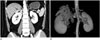
A 49-year-old female presented with a complained symptom of upper respiratory infection, and a contrast-enhanced chest CT scan was done. The CT scan incidentally revealed a tortuous vascular structure in the right suprarenal space. For further evaluation, a contrast-enhanced abdominal CT scan was done (64-section scanner, Lightspeed VCT; GE Medical Systems, Milwaukee, WI, USA) with two phases (corticomedullary and nephrogenic phases, intravenous injection of Iopamidol; Pamiray, Dongkuk Pharm., Seoul, Korea). The tortuous vessels were intertwined with each other around the right adrenal gland (Fig. 1A). The degree of contrast enhancement was similar to that of the abdominal aorta, and we thought it was an arterio-venous malformation. The enhancement pattern of both renal parenchyma was normal, and no hydronephrosis was observed in either kidneys.
The anterior volume-rendered image from CT data (Fig. 1B) showed the tortuous vascular structure at the right renal hilum and the right suprarenal space. Bilateral normal renal arteries and the left renal vein were seen, but the right renal orthopedic vein was not seen in the reconstructed image.
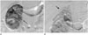
To confirm the arterio-venous malformation and to ensure the proper treatment, we decided to perform an angiography. We punctured the right common femoral artery and selected the right renal artery via a cobra catheter (Cook, Bloomington, MN, USA). The right renal angiogram showed normal contour of a single renal artery and normal renal parenchymal perfusion. After staining the right renal parenchyma, the tortuous vascular structure with small diameter appeared after the renal parenchyma had been stained (Fig. 2A). Then, the suprarenal tortuous vascular structure with large diameter appeared in the delayed phase (Fig. 2B). This tortuous vascular structure continued inferiorly to the level of right renal hilum and ultimately drained into the inferior vena cava (IVC). In the right renal hilum, there was no staining of the normal right renal vein (orthotopic vein) at its expected location. The angiogram finding showed an absence of the orthotropic renal vein and an aberrant formation of suprarenal collateral venous flow.
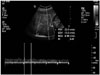
For the confirmation of the venous flow, a Doppler ultrasound (5-8 MHz Curved array probe of iU22 xMATRIX Ultrasound System, Philips, Bothell, WA, USA) was performed. The ultrasound showed an intertwined anechoic tubular structure in the right suprarenal space below the liver capsule. The color Doppler ultrasound revealed vascularity, and the monophasic waveform with a peak velocity of 23.4 cm/s was shown by the spectral Doppler ultrasound (Fig. 3). The suprarenal anechoic tubular structure continued to the renal hilum and showed a unilateral biphasic waveform, which was efferent from the right kidney with a very low resistance (data not shown). These findings confirmed that the vascular structure was composed of dilated veins which drained into the IVC. The peak velocities and resistive index of the right renal artery were normal.
From the CT, angiographic, and ultrasonographic findings, we concluded that there was a developed venous drainage from the right kidney due to the absence of the right orthotopic renal vein. The serum creatinine and BUN levels of the patient were normal. A regular follow-up for the patient was planned.
DISCUSSION
During the embryonic period, both right and left renal veins are formed by the same pattern-anastomosis of the supracardinal and subcardinal veins (1). Anomalies of renal veins have been investigated by many studies, and the most frequent congenital anomaly is an additional renal vein, which is more frequently found on the right side (2). The additional renal vein results in a separate venous drainage of the kidney from the main renal vein, and the flow goes into the IVC independently (5). Compared to the additional renal vein, the absence of a renal vein is a very rare finding, and only two cases have been reported in the English literature (3, 4).
Bozlar et al. (3) reported the aplasia of a right renal vein with a diverted venous drainage, which was a very similar finding to our case. As in our case, any fibrous bands or remnant stalks were not observed at the expected renal vein level of the IVC, and there was no evidence of the parenchymal damage in the right kidney (3). The researchers suggested that a prenatal insult of the renal vein might be the cause of the development of the aberrant venous drainage, prior to the damage of renal parenchyma, which formed the venous varicosity. This explanation could also be applied to our case. Another case of the absence of the right renal vein had a venous varicose complex, which drained into the right gonadal (ovarian) complex (4). We suggest that our case may be similar to venous collateral complex, which developed during embryogenesis.
Unlike the left renal vein, the right renal vein has less prominent collateral flow. In the left renal vein, several venous collateral complexes such as the lumbar veins and the retroaortic and prevertebral venous plexuses are commonly found (6). In an anatomical variation of the right renal vein, the additional renal vein is more commonly found than in the left renal vein (3). Our hypothesis is that congenital or prenatal insult of the right main renal vein would have created the pre-existing additional right renal vein development, which finally became the only aberrant right renal vein. Also, we hypothesize that if a prenatal insult to the left renal vein occurred, the venous collateral complexes of the left renal vein would develop; but, those collateral flows may not have been sufficient for the normal development of the left kidney, in contrast to the additional vein of the right kidney.
Other than the absence of the renal venous drainage, as in our case, there are many acquired causes of renal vein occlusion, such as IVC thrombosis with secondary involvement of the renal vein, hypovolemia, primary renal disease, thrombosis, and trauma (7). Renal vein thrombosis is one of the common causes of renal vein occlusion, which presents with insidious symptoms or frequently without any symptoms (7). The well-established renal venous collateral pathways, other than the main renal vein, take some part of the venous drainage. But, the occluded main renal venous drainage gradually causes renal parenchymal fibrosis histologically, and the other kidney with preserved venous drainage makes up for the abnormal function of the kidney (8). However, our patient had no clinical symptoms or any abnormal lab findings of renal dysfunction, including blood urea nitrogen and serum creatinine. The Doppler ultrasound of the aberrant renal vein showed normal venous drainage into the IVC. Thus, we can assume that the developed aberrant venous drainage could compensate for the absence of the main venous drainage without causing kidney dysfunction. In conclusion, we report an absence of the orthotopic right renal vein with an aberrant renal venous drainage system.
 XML Download