

 PDF
PDF ePub
ePub Citation
 Print
Print


Abstract
Adenoid cystic carcinoma (ACC) is a subtype of adenocarcinoma that is usually seen in the salivary glands. It has also been reported in other organs including the breast, skin, tracheobronchial tree, cervix, larynx, and Bartholin gland. ACC in the breast is rare, accounting for less than 0.1% of all breast cancers. Furthermore, the imaging characteristics of ACC of the breast have not been well described in the literature, especially regarding the findings with magnetic resonance imaging (MRI). Here, we report radiologic findings of a rare case of ACC in the breast by mammography, sonography, computed tomography (CT), positron emission tomography/CT, and MRI with pathologic correlation and a review of the literature.
Figures and Tables
Fig. 1
51-year-old woman with adenoid cystic carcinoma in left breast.
A, B. Craniocaudal and mediolateral oblique views of mammography show a circumscribed round isodense mass in palpable site of the left breast upper center without suspicious calcification.
C, D. Transverse sonography shows about 2.0 cm sized indistinct oval hypoechoic mass in left breast upper center without increased vascularity and posterior features.
LCC = left craniocaudal, LMLO = left mediolateral oblique
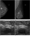
Fig. 2
MRI findings of adenoid cystic carcinoma in the breast.
A. The axial T1-weighted pre-contrast image shows a low signal intensity mass in the left breast.
B. The sagittal T2-weighted image shows a smooth round moderately high signal intensity mass in left breast.
C. The axial dynamic-enhanced T1-weighted image of the first post-contrast acquisition shows about 2.3 cm sized rapid enhancing mass in the left breast.
D. The subtraction image shows persistent enhancement pattern (type I).
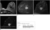
Fig. 3
On the axial positron emission tomography/computed tomography fusion image and MIP, the mass shows hypermetabolism (maximum standardized uptake value: 3.4) without metastasis (arrow).
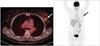
Fig. 4
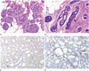
Microscopic findings of pathologically confirmed adenoid cystic carcinoma.
A. The mass is mostly consisted of small basaloid cells. The tumor cells surround pseudoglandular spaces forming variable sized cribriform nests (arrow) (H&E stain, × 100).
B, C. The pseudoglandular spaces are filled with PAS + mucinous materials (arrow) and surrounded by basement membrane materials highlightened by immunohistochemical staining for laminin (B. PAS stain, × 200, C. Laminin, × 100).
D. The tumor cells are focally positive for c-kit (c-kit, × 100).
H&E = hematoxylin and eosin, PAS = Periodic Acid-Schiff
References
1. Santamaría G, Velasco M, Zanón G, Farrús B, Molina R, Solé M, et al. Adenoid cystic carcinoma of the breast: mammographic appearance and pathologic correlation. AJR Am J Roentgenol. 1998; 171:1679–1683.
2. Rosen PP. Adenoid cystic carcinoma of the breast. A morphologically heterogeneous neoplasm. Pathol Annu. 1989; 24(Pt 2):237–254.
3. Lee MS, Kim MK, Kim EK, Park BW, Oh KK. Adenoid cystic carcinoma of the breast: a case report. J Korean Soc Ultrasound Med. 2005; 24:199–202.
4. Glazebrook KN, Reynolds C, Smith RL, Gimenez EI, Boughey JC. Adenoid cystic carcinoma of the breast. AJR Am J Roentgenol. 2010; 194:1391–1396.
5. Youk JH, Kim MJ, Kim EK, Lee JY, Oh KK, Park BW. Recurrence of adenoid cystic carcinoma in the breast after lumpectomy and adjuvant therapy. J Ultrasound Med. 2006; 25:921–924.
6. Tsuboi N, Ogawa Y, Inomata T, Nishioka A, Yoshida D, Yoshida S, et al. Dynamic MR appearance of adenoid cystic carcinoma of the breast in a 67-year-old female. Radiat Med. 1998; 16:225–228.
7. Okamoto Y, Sumiyama Y, Arima Y, Sakuta M, Okuda T, Noto Y, et al. A case of adenoid cystic carcinoma (ACC) of the breast and review of the utility of preoperative imaging diagnose. Breast Cancer. 2001; 8:84–89.
8. Santamaría G, Velasco M, Bargalló X, Caparrós X, Farrús B, Luis Fernández P. Radiologic and pathologic findings in breast tumors with high signal intensity on T2-weighted MR images. Radiographics. 2010; 30:533–548.
9. Ellis IO, Schnitt SJ, Sastre-Garau X, Bussolati G, Tavassoli FA, Eusebi V, et al. Invasive breast carcinoma. In : Tavassoli FA, Devilee P, editors. Pathology and genetics of tumours of the breast and female genital organs. 3rd ed. Lyon, France: IARC;2003. p. 13–59.
10. Kim M, Lee DW, Im J, Suh KJ, Keam B, Moon HG, et al. Adenoid cystic carcinoma of the breast: a case series of six patients and literature review. Cancer Res Treat. 2014; 46:93–97.
 XML Download