

 PDF
PDF ePub
ePub Citation
 Print
Print


INTRODUCTION
Carcinoma of the ampulla of Vater (AoV) is a relatively rare neoplasm, comprising 15-37% of surgically resected pancreatoduodenal tumors and 0.2% of routine autopsy cases; and the most common tumors affecting the AoV are adenocarcinomas (1, 2). Signet ring cell carcinoma (SRCC) of the AoV is rare, and only 21 cases have previously been described in the English language literature (3). Most of the previously reported articles have been published in the clinical or pathology literatures, and little attention has been paid to radiological features of SRCC of the AoV. Here, we present a case of SRCC in the AoV, with the emphasis on imaging features.
Case Report
A 41-year-old man was hospitalized for 20 days with jaundice. Upon physical examination, he presented with icteric sclera and visible jaundice. Liver function tests showed total bilirubin 7.5 mg/dL, aspartate aminotransferase 142 IU/L, alanine aminotransferase 159 IU/L, alkaline phosphatase 420 IU/L, and γ-glutamyl transferase 1048 IU/L. Tumor marker, carbohydrate antigen 19-9 was elevated (53.6 U/mL). The urine color was amber and contained 3.0 mg/dL of bilirubin.
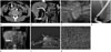
Dynamic contrast-enhanced 64-channel multidetector computed tomography of the liver showed a 1.2-cm sized lobulating contoured mass with enhancement in the AoV. The upstream intrahepatic and extrahepatic bile ducts were proportionally dilated (Fig. 1A, B). These image findings suggested a carcinoma of the AoV with direct invasion of the distal common bile duct (CBD). There was no evidence of distant metastases. Endoscopic retrograde cholangiopancreatography revealed an ulcerofungating mass in the AoV. Diffuse dilatation of the bile duct was noted, and there was a focal nodular contrast filling defect in the far-distal CBD (Fig. 1C, D). Dilated bile duct was decompressed with endoscopic nasobiliary drainage (ENBD). Endoscopic retrograde cholangioscope-guided biopsy samples from the mass of the AoV showed moderately differentiated adenocarcinoma. Gadoxetic acid (Gd-EOB-DTPA, Primovist; Bayer HealthCare, Berlin, Germany) enhanced magnetic resonance cholangiography revealed a distinct nodular enhancing lesion in the AoV similar to CT, and bilary system was well decompressed by ENBD. The 18-fluoro-2-deoxy-D-glucose positron emission tomography/computed tomography (18F-FDG PET/CT) showed increased FDG uptake (maximum standardized uptake value 4.1) in the distal CBD (Fig. 1E).
The patient underwent a pylorus preserving pancreaticoduodenectomy. On gross evaluation of the resected specimen, mucosal surface of the duodenum showed a polypoid mass with central ulceration, measuring 2.0 × 1.6 cm in diameter in the AoV. The cut surface of the ampullary tumor revealed a whitish tumor with pushing margins, measuring 1.1 × 1 cm in diameter. The tumor invaded through the duodenal wall but the pancreas was grossly unremarkable (Fig. 1F). The histology of tumor was composed of usual signet ring cells, and the lower half was consisted of eosinophilic signet ring cells (Fig. 1G). As a result of these findings, the final histopathologic diagnosis was SRCC. The tumor cells were found in two regional lymph nodes. According to the TNM classification, tumor of the AoV was T2N1M0 (Stage IIB).
After the surgery, the patient underwent five cycles of postoperative chemotherapy with combination of 5-fluorouracil and cisplatin chemotherapy regimen. On the follow-up CT scans, local recurrence or metastatic disease were not detected. The patient was alive and disease-free at the 13 months post-operative follow-up.
DISCUSSION
Carcinoma of the AoV is a relatively rare neoplasm, and the most common tumors affecting the AoV are adenocarcinomas (1, 2). Ampullary carcinomas typically manifest as small tumors at the time of diagnosis, because of the relatively early onset of symptoms; therefore, the mass itself is often not apparent on imaging. However, secondary findings such as marked bile duct dilatation, in association with mild to moderate dilatation of the pancreatic duct, can usually be seen on CT. Large ampullary tumors usually manifest as an infiltrative or polypoid mass (4).
SRCC can arise in many organs, but it usually occurs in the gastrointestinal tract, especially in the stomach. It has been reported that 90% of SRCC occurs in the stomach, with the rest arising in several other organs, including the breast, gallbladder, pancreas, urinary bladder, and colon (5). When it arises from the stomach, SRCC shows stronger enhancement than non-signet ring cell carcinoma (6) and is generally known for its advanced stage and poor prognosis (7). SRCC of the biliary system is rare and most SRCCs of the biliary system are originated from gallbladder (8). Because of its rareness, little is known about the origin of SRCCs in the periampullary area, and two possible explanations for this histologic variation were suggested. One explanation is that the tumors may arise from ectopic gastric mucosa. Indeed, there are some reported cases of SRCCs with ectopic gastric mucosa in the ampullary tumors. Another theory holds that these carcinomas arise from the areas of gastric-type metaplastic epithelium, which are considered to be a protective response to elevated acidity and are observable in the duodenal bulb of patients with peptic ulcer patients (7, 8).
Several modalities such as CT, MRI, and US (including contrast-enhanced US) were used to diagnose the SRCC of the AoV in the previously reported cases. All accessible literatures only showed dilated bile ducts without an apparent mass lesion in the AoV, which are unable to reveal malignant etiology, except for Gao et al. (9) who reported with contrast-enhanced US. Unlike these previously reported cases, we were able to detect the tumor.
According to Chang et al. (10), mean CT attenuation values of ampullary tumor was 107 ± 22 Hounsfield unit (HU) (in the arterial phase) and 103 ± 17 HU (in the portal venous phase). In our case, the tumor showed 120 HU and 148 HU, respectively. The attenuation numbers were higher than average ampullary tumor. This enhancement pattern is similar to the gastric SRCC pattern (6). But more cases are needed to confirm hyperenhancement as a feature of SRCC of the AoV, since this is the first case to describe it. In previously reported 21 cases, only two cases had lymph node metastasis at the diagnosis, and this presenting case also had two metastatic lymph nodes. The nodes were neither significantly enlarged on CT nor showed increased FDG uptake on PET/CT.
In conclusion, little is known about the imaging features of SRCC in the AoV; but considering the poor prognosis of SRCCs of other sites, radiologists and clinicians should be aware of the possibility of a SRCC when hyper-enhancing tumor in the AoV is encountered.
 XML Download