

 PDF
PDF ePub
ePub Citation
 Print
Print


INTRODUCTION
The common femoral artery is one of the most common access routes for endovascular treatment. Manual compression has been the traditional mainstay for hemostasis of the femoral arteriotomy and remains the "gold standard" (1). However, various arterial closure devices (ACDs) have been developed, given the prolonged bed rest of patients following during manual compression. These devices utilize either a collagen plug or sutures to control bleeding (2, 3, 4, 5). In contrast, mechanical compression devices (MCDs) have been designed to replace or assist with manual compression, and most available disposable devices (Femostop Gold, St. Jude Medical, St. Paul, MN, USA; Safeguard, Maquet, Rastatt, Germany; and GH-150, Kyung-Won Medical, Seoul, Korea) use a pneumatic system to deliver pressure (6, 7, 8). It is reasonable to assume that MCDs have a possible advantage over ACDs, as no foreign body is left in the body; therefore, repeated punctures may be tolerated more easily. In this study, the safety and hemostatic efficacy of a novel MCD, which utilizes directly applied pressure with a non-compliant disk (Xpress, Angiovention, Seoul, Korea), was analyzed retrospectively.
MATERIALS AND METHODS
Patients
All patients included in this study signed written informed consent for use of the Xpress device at the time of the procedure. Our institutional review board waived additional informed consent for this retrospective study. Our interventional radiology database search revealed 290 consecutive patients from October 2013 to January 2014, in whom a femoral arteriotomy site was post-procedurally compressed using the Xpress device to achieve hemostasis. The Xpress device was routinely used in our department for the 391 consecutive patients who underwent a transfemoral arterial intervention during the period. Patients with a high risk of developing hemorrhage were compressed manually by a physician, followed by applying the Xpress device. Among those who elected to use an ACD, Xpress was used selectively when the ACD failed. Otherwise, all patients received the Xpress device as the sole means for hemostasis without manual compression.
The inclusion criteria for the study population were: 1) patients > 18 years and 2) use of the Xpress device without concomitant ACD use or manual compression. Patients were excluded only if they had incomplete medical records regarding the femoral arteriotomy site. A total of 290 patients met the inclusion criteria. Among them, 39 were excluded because the post-procedural medical records regarding the femoral arteriotomy site were incomplete. Finally, 251 patients were included in the study.
The patient characteristics and periprocedural factors are summarized in Table 1.
Medical Record Review
The patient electronic medical records were reviewed retrospectively,
including the following factors: age, sex, body mass index (BMI), pre-procedural platelet count, prothrombin time international normalized ratio, activated partial prothrombin time, systolic and diastolic blood pressure, heart rate at the time of hemostasis, procedure duration, vascular sheath size used, and time to apply the compression device.
Device Application
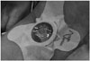
The Xpress device is composed of a rigid plastic dome-shaped disk with a rotator handle to adjust pressure, which is attached to a quadrilobed adhesive band. A separate focus pad is provided, and may be optionally applied to the puncture site to increase pressure in obese patients. Two additional sterile adhesive bands are also included in the package to enhance compression. The exact pressure applied to the arteriotomy site is not quantifiable by the device; therefore, it is adjusted by the physician based on stopping the hemorrhage and preserving the dorsalis pedis arterial pulse. Mild pain was mostly transient, and oral analgesics were prescribed occasionally.
The vascular sheath was pulled out about 3 cm at the end of each endovascular procedure, and the device was applied with the pressure disk placed at the center of the arteriotomy site. Then, clock-wise rotation was applied to compress the puncture site. Subsequently, the vascular sheath was removed gently. Possible hemorrhage or swelling of the inguinal area was observed through a transparent window with hands off the device (Fig. 1). If persistent bleeding was observed, the additional adhesive bands included in the device package were applied to increase pressure at the arteriotomy site. The patients were observed in the angiography room for the first 30 min and were then sent to the ward or intensive care unit where the nursing staff was instructed to keep the devices in place for at least 3 hours. Hemostasis was determined by the nursing staff or a radiology resident when the rigid disk of the device was decompressed. The devices were removed when hemostasis was achieved or the femoral arteriotomy site was recompressed for an additional 30 min using the device.
Definition of Successful Hemostasis
Successful immediate hemostasis was defined as complete cessation of bleeding from the arteriotomy site without inguinal swelling during the first 30 min after applying the device. Successful delayed hemostasis was defined as complete cessation of bleeding without minor or major complications for 4 hours. Major and minor complications were classified according to the Society of Interventional Radiology guidelines (9).
Statistical Analysis
Immediate and delayed hemostatic rates were calculated with 95% confidence intervals (CIs) using the Pearson-Klopper method. The patients were further divided into complication-free and complication groups (patients with either minor or major complications). The two groups were compared using the χ2 test for continuous variables, and Student's t-test for binary variables. A p-value < 0.05 was considered significant. All statistical analyses were performed using the R: A Language and Environment for Statistical Computing, ver. 3.1.1 software (R Foundation for Statistical Computing, Vienna, Austria).
RESULTS
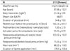
Successful immediate hemostasis was achieved in 250 of 251 patients (99.6%; 95% CI, 97.8-100.0%). Successful delayed hemostasis was achieved in 236 of 251 patients (94.0%; 95% CI, 90.3-96.6%). One patient, who failed immediate hemostasis, was managed with manual compression and was considered a technical failure.
No major complications were detected, and all complications were minor. Eight patients (3.19%; 95% CI, 1.4-6.2%) developed minimal oozing, and six patients developed small inguinal hematomas < 5 cm in size (2.39%; 95% CI, 0.9-5.1%), which were managed with a sandbag applied to the puncture site.
The subgroup analysis between the complication-free and patients with complications is summarized in Table 2. Complications were more prevalent in younger patients (p = 0.014). The duration of compression by the device was significantly shorter among patients who developed complications (p = 0.007). Larger vascular sheath size was associated with the development of complications (p = 0.002).
DISCUSSION
Hemostasis of a femoral arteriotomy site can be achieved using manual compression with or without a topical patch, MCD, or ACD (1, 10, 11). All three ways have different mechanisms; therefore, they vary in terms of efficacy and safety, and disadvantages should be expected.
In general, ACDs include suture-mediated and collagen-plug based types. A few long-term studies have reported the safety of ACDs. A meta-analysis of randomized trials on the efficacy of ACDs compared to that of manual or mechanical compression reported a significantly increased incidence of groin infection (0.20% vs. 0.06%) after using ACDs (12). In contrast, ACDs facilitate hemostasis; thus, allowing early ambulation.
Another recently published meta-analysis revealed marginally fewer complications with ACDs compared with manual compression, but the difference was not statistically significant (13). Another study found no differences in complication rates among patients treated for femoral hematomas, pseudoaneurysms, or distal ischemia by manual compression and an ACD (13). A possible explanation for the discrepancy is that there are only a few robust clinical trials regarding femoral hemostasis.
Koreny et al. (10) reported a 2004 systematic review that was based on 30 randomized trials. The results showed no significant differences in relative risk (RR) in patients treated for groin hematoma, pseudoaneurysm, or arteriovenous fistula with an ACD compared with those treated by manual compression. However, when the analyses were limited to studies of good methodological quality, ACDs were associated with higher risks for hematoma (RR, 1.89; 95% CI, 1.13-3.15) and pseudoaneurysm (RR, 5.40; 95% CI, 1.21-24.5). Similarly, Nikolsky et al. (14) reported in 2004 that a specific type of ACD (VasoSeal) was associated with an increased risk of complications during percutaneous coronary intervention, but did not show a significant difference in terms of complication risk, compared with that of manual compression in a diagnostic setting.
These striking differences regarding complications after applying an ACD compared with those following manual compression may be largely operator dependant. Another possible explanation is that ACDs have been improved during the last decade, and later studies have shown better results with ACDs. A 2007 analysis of a prospective registry reported that routine use of ACDs decreases the risk of vascular complications following diagnostic coronary catheterization by 58% (95% CI, 19-88%) and 42% (95% CI, 17-59%) following percutaneous coronary intervention (15).
However, most available MCDs use a pneumatic system to achieve hemostasis of a femoral arteriotomy site (6, 7, 8). Among them, Femostop is one of the most widely studied devices and results in fewer complications than those from manual compression (7).
The XPress device is rarer among femoral compression devices. It uses direct external pressure with a rigid noncompliant disk rather than pneumatic pressure to facilitate hemostasis, and no foreign material is left in the femoral artery. The Xpress device is unique in design, as it has a noncompliant disk used to compress the arteriotomy site, while most other MCDs use a pneumatic system. The Xpress device delivers constant pressure over the arteriotomy site compared to that of manual compression. A few other commercial devices are available with a similar hemostatic mechanism. Femostop is one of the more studied among the femoral compression devices. Major complication rates for the Femostop device are 1.9-13.9% (7, 8, 16). The complication rate in our study was comparable to that of previous compression devices and is acceptable according to recently published guidelines advising that the complication rate should be < 3% (1).
A long-term observational study with a mean follow-up of 3320 days in patients who underwent coronary artery intervention showed that femoral ACDs did not significantly affect distal blood flow through the femoral arteriotomy site (17). However, there is a paucity of data on femoral arteries that have been repeatedly punctured and received an ACD. ACDs generally leave foreign material in the body, whereas MCDs do not. MCDs substitute for physician compression with a machine, and nothing is left in the body. Therefore, it is reasonable to use a MCD in a patient who is expected to undergo multiple trans-femoral intervention sessions.
Our results showed no major complication following use of this new MCD. One possible reason for the absence of major complications in our study was that our patients had low BMIs (23.74 ± 2.91 kg/m2); thus, it was easier to stop bleeding by manual compression. Another reason for the lower complication rate in our study could be selection bias from the retrospective design. We opted to use suture-mediated ACDs to minimize bleeding complications in patients who were expected to bleed.
Interestingly, our results reveal that significantly more young patients developed minor complications. We hypothesize that younger patients were more likely to be active, and that these patients may have been less compliant with the physician's instruction not to move their legs during bed rest.
As expected, vascular sheath size impacted the development of complications in our study population. Patients who received larger sheath sizes were more likely to suffer complications.
This study had several limitations. First, it was designed retrospectively, and use of a MCD was at the discretion of the interventionalist; therefore, selection bias was unavoidable. We routinely use MCD devices unless the patient or referring physician requests an ACD, or the patient refuses use of a MCD. Second, most patients had low BMIs, which lead to a greater hemostatic success rate using external compression. Third, we did not compare other hemostatic devices or manual compression. Last, no cost-effectiveness analysis was conducted. Notably, given the simple device design with a noncompliant plastic disk and adhesive tape, the cost of the device in our hospital is around $50 USD, which is lower than that for a combined ACD and MCD in our hospital.
In conclusion, our results showed an excellent hemostatic rate with use of the Xpress device in patients with a relatively low BMI. Further studies comparing the Xpress device with other ACDs, MCDs, and manual compression, as well as analyzing cost-effectiveness are necessary to confirm this initial experience.

 XML Download