

 PDF
PDF ePub
ePub Citation
 Print
Print


INTRODUCTION
Large or giant aneurysms in the cavernous segment of the internal carotid artery (ICA) frequently present with neurological symptoms caused by the mass effect on the cranial nerves. Thus, the treatment goal for a symptomatic giant ICA aneurysm in the cavernous segment is resolution of the mass effect that leads to cranial nerve dysfunctions (1). Several endovascular treatments for giant aneurysms have already been introduced, such as detachable coiling and flow diverter stent insertion. However, complete packing of the aneurismal sac itself is difficult, and the recurrence rate is high (2). These endovascular techniques can also cause transient swelling of the aneurysmal sac after induced thrombosis as well as delayed complications, including bleeding. In this case report, we described our first use of surgical n-butyl 2-cyanoacrylate (NBCA) and coils for treating a symptomatic, giant saccular aneurysm located in the cavernous segment of the ICA.
CASE REPORT
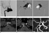
A 52-year-old female presented with tinnitus and intermittent diplopia. A cerebral angiography showed a giant aneurysm (31.4 × 15.7 × 17.5 mm) arising in the cavernous segment (C4, ascending segment) and protruding medially (Fig. 1A). An initial magnetic resonance imaging (MRI) showed a partially thrombosed, giant aneurysm at the level of the right cavernous sinus and protruding into the sphenoid sinus. The patient underwent a balloon occlusion test of the right ICA for 20 minutes, and there was no focal neurologic sign or lateralizing sign seen during the occlusion test. A single photon emission CT using 99mTc hexamethylpropyleneamineoxime injected during the balloon inflation demonstrated no cerebral perfusion alteration.
Following the balloon occlusion test, the right ICA was trapped by coiling at two points, i.e., just proximal and distal to the aneurysm. After isolating the aneurysm with a complete ICA occlusion, a 25% NBCA-lipiodol mixture was injected into the aneurismal sac in order to prevent volume expansion by thrombosis (Fig. 1B, C), and additional coil packing at the petrous and cervical ICA was done. Finally, stump occlusion using a 6 mm Amplatzer vascular plug was done in the cervical ICA.
Immediately after the procedure, the patient developed partial sixth nerve palsy, presenting with mild diplopia, which was completely resolved with an anti-inflammatory medication. A follow-up MRI done 15 months later showed the decreased size of the thrombosed aneurysm in the source magnetic resonance angiography (MRA) image compared to the initial MRA image (Fig. 1D, E). There was good filling of the ipsilateral cerebral vessels with an obliteration of the right ICA and the aneurysm (Fig. 1F). The patient did not express any complaints and did not reveal any neurological deficit during the follow-up period.
DISCUSSION
Many giant aneurysms present with symptoms caused by their mass effect. The exact clinical manifestation of the mass effect depends on the location of the aneurysm and the involved adjacent neural structures. Giant aneurysms arising from the cavernous and proximal intracranial carotid arteries present with dysfunction of ocular movement as well as vision deterioration (3, 4). During the past 30 years, several endovascular treatments for giant cavernous ICA aneurysm have been introduced, such as detachable coiling and flow diverter stent insertion. Detachable coils can be used for packing of the aneurismal sac itself or of the parent artery (5). Flow diverter stents are used to restrict and redirect blood flow along the vessel axis for reconstructing the parent vessel lumen across the aneurysm neck, thus promoting aneurysm thrombosis (6).
These endovascular techniques can, however, lead to unfavorable clinical outcomes, including ischemia and compression syndromes caused by transient aneurysm swelling after an induced sac thrombosis and subsequent bleeding of a previously un-ruptured aneurysm (7). In addition, the incidence of recanalization and incomplete embolization of the giant aneurysm lumen is high due to the difficulty of compact coil packing for the large volume (2).
In order to obliterate dead space in the aneurysmal sac, which is subject to being thrombosed, we used a 25% NBCA-lipiodol mixture after coil packing of the parent arterial lumem. We expected that the penetration of glue in the aneurysm could prevent it from migrating distally through the coil mesh in the completely occluded distal ICA lumen. In vitro studies of the 28% glue concentration in a previous study, sticking of the micro-catheter to the glue cast did not occur (8). In addition, there was no resistance during the retrieval of the micro-catheter.
The aneurysm size may decrease after NBCA embolization, as seen on the follow-up MRI. It seemed to result from three postulated mechanisms as follows (8, 9): 1) NBCA in the aneurysm is absorbed by the blood; 2) NBCA in the aneurysm is absorbed through the aneurysmal wall after an inflammatory process caused by the NBCA; or 3) there is extra-luminal migration of the NBCA through the aneurysmal wall. On this basis, the transient diplopia that occurred in our patient can be explained by an inflammatory process in the aneurysmal wall, with the use of 25% concentration NBCA. A future study to find out the exact mechanism of this phenomenon is required. We believe that the decrease in the aneurysm size after a NBCA embolization can result in a mass-relieving effect, which can also be achieved by a parent artery occlusion (10).
Our report has several limitations for further considerations. First, we could not completely obliterate the mass effect using NBCA, even though the mass effect was not serious. Therefore, it would be necessary to both predict and manage any further mass effect by using steroids, for example. Secondly, the injected low concentration of NBCA slowly had contact with the blood in the aneurysm and slowly spread the coagulation effect during the real-time fluoroscopy, thus suggesting a potential effect of volume expansion. Therefore, a slow injection of NBCA is required with careful real-time fluoroscopic observation of the direction and extent of the NBCA.
 XML Download