

 PDF
PDF ePub
ePub Citation
 Print
Print


INTRODUCTION
Pseudoaneurysm, also known as false aneurysm, is a hematoma resulting from a defect in the vascular wall that freely communicates with the parent vessel. The most common causes of massive lower gastrointestinal (GI) bleeding are diverticular disease, angiodysplasia, and colitis, whereas pseudoaneurysmal lower GI bleeding is not common. We report a rare case of traumatic superior rectal artery pseudoaneurysm presenting with recurrent lower GI and pelvic extraperitoneal bleeding resulting from a penetrating perineal injury.
CASE REPORT
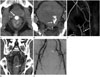
A 43-year-old male patient who was a chronic alcoholic, presented in a drunken state with a penetrating perineal wound, caused by a wooden stick and resulting in massive lower GI bleeding. Computed tomography (CT) at a local hospital on the day of trauma showed an abnormal enhancing saccular lesion at the anterior wall of the rectum, 6 cm proximal from the anal verge and extending upward to the level of the bladder neck. The lesion was thought to be supplied by the superior rectal artery from the inferior mesenteric artery and was suggestive of a pseudoaneurysm (Fig. 1A). The pseudoaneurysm was saccular in shape and surrounded by a small hematoma in the posterior perivesicular space. The patient was managed at a local hospital, where he underwent an emergent laparoscopic Hartmann procedure consisting of diversion sigmoid end colostomy with a sutured rectal stump left behind without resection of the pseudoaneurysm.
The patient had secondary lower GI bleeding after surgery on the same day. The perineal wound and rectum were packed with gauze and the patient was referred to our hospital on day 2 of trauma. The patient arrived with a blood pressure of 100/40 mm Hg and a tachycardia of 141 beats per minute. He had abdominal distention and his abdomen was vaguely guarded. The hemoglobin level was 8.2 g/dL on arrival. The patient was presumptively diagnosed as rupture of the Hartmann pouch (rectal stump). On day 2 of trauma, follow-up CT demonstrated a large 32hematoma due to rupture of the pseudoaneurysm and active contrast leakage from the pseudoaneurysm (Fig. 1B, C). On day 3 of trauma, the patient became hemodynamically unstable and had emergent revision Hartmann procedure and hematoma evacuation because of unknown pseudoaneurysm. On day 4 of trauma, follow-up CT showed almost complete clearing of the pelvic hematoma but a small focal enhancing lesion was noted espein the anterior wall of the rectum. We diagnosed this as a partially thrombosed residual pseudoaneurysm at the anterior wall of the rectum (Fig. 1D).
On day 14 of trauma, the patient's blood pressure had dropped to 100/50 mm Hg and hemoglobin level was decreased to 7.9 g/dL. Internal bleeding from ruptured pseudoaneurysm was suspected; this was the third bleeding episode. A percutaneous drainage catheter was temporarily inserted into the abdominal cavity. On day 17 of trauma, the patient became hemodynamically stabilized. CT showed a pelvic extraperitoneal hematoma, probably representing bleeding from the residual pseudoaneurysm, but active contrast leakage was not evident (not shown). On day 19 of trauma, angiography was performed to treat the residual pseudoaneurysm. A conventional angiography via the right common femoral artery displayed no pseudoaneurysm of the superior rectal artery or extravasation from either bilateral internal iliac arteries or inferior mesenteric artery (Fig. 1E). Therefore, we performed prophylactic Gelfoam embolization of the inferior mesenteric artery and the anterior branch of bilateral internal iliac arteries. We concluded that spontaneous healing of the pseudoaneurysm had occurred. On day 31 of trauma, follow-up CT showed a decreasing pelvic hematoma and no evidence of residual pseudoaneurysm. The patient recovered well and no further episodes of lower GI bleeding have been reported. After 8 weeks, the sigmoid colostomy was reversed.
DISCUSSION
Pseudoaneurysm is a vascular out-pouching from the parent artery and lacks a complete arterial wall. Factors such as trauma, inflammation, or infection are known causes of pseudoaneurysm. Complications of pseudoaneurysm include early rupture, compression of adjacent structures, and combined infection (1). Early diagnosis of pseudoaneurysm is important because attempts at post-rupture surgical repair lead to a high mortality rate of 50% (2). Ultrasonography, CT, and magnetic resonance imaging enable detection of visceral vascular lesions, but conventional angiography is important for further diagnosis and treatment. Endovascular treatment is often the first-line therapy. Endovascular intervention or open surgical repair is necessary for all pseudoaneurysms (1).
To our knowledge, there are 3 case reports in the literature regarding massive lower GI bleeding resulting from a superior rectal artery pseudoaneurysm. Janmohamed et al. (3) reported a case of lower GI bleeding caused by non-traumatic pseudoaneurysm secondary to acute diverticulitis, which was diagnosed by CT angiography. Our radiologic finding was saccular pseudoaneurysm, similar to the previously reported case. The other two cases (4, 5) were traumatic pseudoaneurysms resulting from penetrating trauma or iatrogenic cause (colonoscopic procedure). All three cases were treated by angiographic embolization. We performed surgical hematoma removal alone without resection of pseudoaneurysm that ruptured thereafter.
Some authors (6,7,8) reported spontaneous occlusion of traumatic pseudoaneurysms resulting from thrombosis within 2 weeks. The pseudoaneurysm in our patient was apparent on CT, but not on angiography after the third bleeding following the revision Hartmann procedure. This was because of the 11 cm segmental resection of the rectosigmoid colon with ligation of the superior and middle rectal arteries. Furthermore, partial thrombosis of the pseudoaneurysm seemed to hinder the opacification of residual pseudoaneurysm and caused spontaneous healing. Yi et al. (9) reported a case of prophylactic embolization of traumatic hepatic artery pseudoaneurysm. Prophylactic embolization is required to prevent severe hemorrhage from pseudoaneurysm rupture. We likewise subsequently performed prophylactic embolization of the inferior mesenteric artery and the anterior branch of bilateral internal iliac arteries to treat the residual pseudoaneurysm.
The patient in the present case had recurrent massive lower GI and pelvic extraperitoneal bleeding that could not be controlled by repeated surgical attempts. It was likely that a surgeon at the local hospital performed an emergent end colostomy on the day of trauma without recognition of the pseudoaneurysm depicted on CT. On day 3 of trauma, the surgeon at our hospital performed an emergent hematoma evacuation and revision end colostomy, again with failure to recognize the pseudoaneurysm on CT performed at our hospital. Additionally, the surgical approach and visualization of the pseudoaneurysm might have been difficult due to its deep position. Hence, there was a residual partially thrombosed pseudoaneurysm, whose rupture resulted in the third bleeding. Vascular lesions such as pseudoaneurysms should be considered in the differential diagnosis of patients with post-traumatic lower GI bleeding, and more especially in cases of penetrating injury, for effective treatments such as complete surgical removal or endovascular exclusion of flow.
In conclusion, we reported a rare case of traumatic superior rectal artery pseudoaneurysm with recurrent lower GI and pelvic extraperitoneal bleeding that was caused by a lack of pretreatment recognition of the pseudoaneurysm. Third bleeding on day 14 of trauma, in particular, could have been prevented if the surgeon had been informed about the pseudoaneurysm in a timely manner. Therefore, the radiologist should be aware of CT imaging findings of pseudoaneurysms and the surgeon should recognize the details regarding the parent vessel, location, and size of the pseudoaneurysm before surgery in select clinical candidates.
 XML Download