

 PDF
PDF ePub
ePub Citation
 Print
Print


Abstract
Purpose
To present the clinical outcome of computed tomography (CT) guided radiofrequency ablation (RFA) for osteoid osteoma.
Materials and Methods
Thirty-one patients (M:F = 23:8, mean age: 20 years, range: 4-54 years) who underwent RFA for clinically suspected osteoid osteoma from May 2004 to December 2013 were retrospectively reviewed. RFA was done in all cases under CT guidance by one of three radiologists in our department. Electronic medical records and images were retrospectively reviewed in all patients.
Results
Lesions were located in femur (n = 20), tibia (n = 5), fibula (n = 2), humerus (n = 3), talus (n = 2), and calcaneus (n = 1). On discharge, 27 of 33 cases showed complete remission of pain (82%). One major complication (compartment syndrome) and 2 minor complications (reactive synovitis, minimal skin burn at electrode insertion site) were observed. On the last follow-up (0-78 months, mean: 12.6 months) 27 of 33 cases were successfully treated (82%) and had no more complaints. 3 cases presented remaining pain (9%). In 3 cases relapse occurred (9%) and RFA was repeated in 1 case. The repeated treatment was successful.
Figures and Tables
Fig. 1
A 14-year-old female patient who was diagnosed as osteoid osteoma.
A. Non-contrast hip-CT shows typical nidus with internal calcification at right distal femur with adjacent prominent cortical thickening (black arrow).
B. MRI shows intermediate signal intensity (SI) in the nidus on T1-weighted image and high SI on T2-weighted image with internal dark SI of calcification on all sequences (white arrows).
C. The lesion was successfully treated by computed tomography guided radiofrequency ablation.
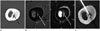
Fig. 2
A 40-year-old male patient who was diagnosed as osteoid osteoma.
A. Axial and coronal hip-CT scan shows a small osteolytic lesion with surrounding cortical thickening and sclerosis at left proximal femur, lesser trochanteric area (white arrows). The lesion is suspected as intra-articular location.
B. To avoid possible heat damage to the surrounding tissue around the hip joint, needle tip is located at deep aspect to the nidus.
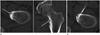
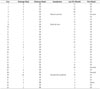
Table 1
Summary of Cases
Table 2
Result
References
1. Frassica FJ, Waltrip RL, Sponseller PD, Ma LD, McCarthy EF Jr. Clinicopathologic features and treatment of osteoid osteoma and osteoblastoma in children and adolescents. Orthop Clin North Am. 1996; 27:559–574.
2. Cohen MD, Harrington TM, Ginsburg WW. Osteoid osteoma: 95 cases and a review of the literature. Semin Arthritis Rheum. 1983; 12:265–281.
3. Boscainos PJ, Cousins GR, Kulshreshtha R, Oliver TB, Papagelopoulos PJ. Osteoid osteoma. Orthopedics. 2013; 36:792–800.
4. Pikoulas C, Mantzikopoulos G, Thanos L, Passomenos D, Dalamarinis C, Glampedaki-Dagianta K. Unusually located osteoid osteomas. Eur J Radiol. 1995; 20:120–125.
5. Assoun J, Railhac JJ, Bonnevialle P, Poey C, Salles de Gauzy J, Baunin C, et al. Osteoid osteoma: percutaneous resection with CT guidance. Radiology. 1993; 188:541–547.
6. Davies M, Cassar-Pullicino VN, Davies AM, McCall IW, Tyrrell PN. The diagnostic accuracy of MR imaging in osteoid osteoma. Skeletal Radiol. 2002; 31:559–569.
7. Parlier-Cuau C, Champsaur P, Nizard R, Hamze B, Laredo JD. Percutaneous removal of osteoid osteoma. Radiol Clin North Am. 1998; 36:559–566.
8. Rosenthal DI, Springfield DS, Gebhardt MC, Rosenberg AE, Mankin HJ. Osteoid osteoma: percutaneous radio-frequency ablation. Radiology. 1995; 197:451–454.
9. Rosenthal DI, Alexander A, Rosenberg AE, Springfield D. Ablation of osteoid osteomas with a percutaneously placed electrode: a new procedure. Radiology. 1992; 183:29–33.
10. Bruners P, Penzkofer T, Günther RW, Mahnken A. [Percutaneous radiofrequency ablation of osteoid osteomas: technique and results]. Rofo. 2009; 181:740–747.
11. Hoffmann RT, Jakobs TF, Kubisch CH, Trumm CG, Weber C, Duerr HR, et al. Radiofrequency ablation in the treatment of osteoid osteoma-5-year experience. Eur J Radiol. 2010; 73:374–379.
12. Kjar RA, Powell GJ, Schilcht SM, Smith PJ, Slavin J, Choong PF. Percutaneous radiofrequency ablation for osteoid osteoma: experience with a new treatment. Med J Aust. 2006; 184:563–565.
13. Schmidt D, Clasen S, Schaefer JF, Rempp H, Duda S, Trübenbach J, et al. [CT-guided radiofrequency (RF) ablation of osteoid osteoma: clinical long-term results]. Rofo. 2011; 183:381–387.
14. Rosenthal DI, Hornicek FJ, Wolfe MW, Jennings LC, Gebhardt MC, Mankin HJ. Percutaneous radiofrequency coagulation of osteoid osteoma compared with operative treatment. J Bone Joint Surg Am. 1998; 80:815–821.
15. Yildiz Y, Bayrakci K, Altay M, Saglik Y. Osteoid osteoma: the results of surgical treatment. Int Orthop. 2001; 25:119–122.
16. Vanderschueren GM, Taminiau AH, Obermann WR, Bloem JL. Osteoid osteoma: clinical results with thermocoagulation. Radiology. 2002; 224:82–86.
17. Rimondi E, Mavrogenis AF, Rossi G, Ciminari R, Malaguti C, Tranfaglia C, et al. Radiofrequency ablation for non-spinal osteoid osteomas in 557 patients. Eur Radiol. 2012; 22:181–188.
18. Cribb GL, Goude WH, Cool P, Tins B, Cassar-Pullicino VN, Mangham DC. Percutaneous radiofrequency thermocoagulation of osteoid osteomas: factors affecting therapeutic outcome. Skeletal Radiol. 2005; 34:702–706.
19. Finstein JL, Hosalkar HS, Ogilvie CM, Lackman RD. Case reports: an unusual complication of radiofrequency ablation treatment of osteoid osteoma. Clin Orthop Relat Res. 2006; 448:248–251.
20. Sung KS, Seo JG, Ha HC. CT-guided Percutaneous Thermoablation for the Treatment of Osteoid Osteoma. J Korean Bone Joint Tumor Soc. 2004; 10:88–95.
21. Lindner NJ, Ozaki T, Roedl R, Gosheger G, Winkelmann W, Wörtler K. Percutaneous radiofrequency ablation in osteoid osteoma. J Bone Joint Surg Br. 2001; 83:391–396.
22. Sim FH, Dahlin CD, Beabout JW. Osteoid-osteoma: diagnostic problems. J Bone Joint Surg Am. 1975; 57:154–159.
 XML Download