

 PDF
PDF ePub
ePub Citation
 Print
Print


INTRODUCTION
Cardiac lipoma is a rare benign primary cardiac tumor and is usually asymptomatic (1). It is usually discovered incidentally but can be related to hemodynamic obstruction depending on its location (2). There have been several reports regarding cardiac lipoma, including its clinical presentation and characteristic findings on computed tomography (CT) (2, 3). However, few cases documented the characteristic CT findings of solitary or multiple cardiac lipomas in the patient with tuberous sclerosis; and to our knowledge, no cases of biventricular lipoma or lipomatous hypertrophy with multifocal fat in a non-tuberous sclerosis patient have ever been reported (4). Therefore, we here report a case of right ventricular (RV) lipomatous mass and multifocal fat infiltration in both ventricles, which were incidentally detected in a young woman showing an abnormal electrocardiogram (ECG) finding.
CASE REPORT
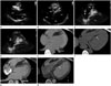
A 31-year-old woman, at 37 + 4 weeks of pregnancy, showed T-wave inversion on the precordial leads on a scheduled ECG, which was part of a preoperative evaluation for Cesarean section. She had a history of myomectomy and a Cesarean section two years ago. The physical examination was normal, and there were no skin lesions found at the admission. There also were no cardiac symptoms and the vital signs were within normal range. The initial blood pressure was 120/80 mmHg, and the respiratory rate was 20 breaths per minute. Her body mass index was 24.97 kg/m2. There was no elevation of cardiac enzymes. Transthoracic echocardiography (TTE) was performed for further evaluation of abnormal ECG findings. On TTE, a hyperechoic soft tissue mass-like lesion was noted on the interventricular septum in the RV (Fig. 1A-C), and another echogenic mass-like lesion was noted adherent to the RV free wall (Fig. 1C). The largest mass measured about 18 × 50 mm in size. However, there was no evidence of hemodynamic occlusion on Doppler echocardiography (Fig. 1D). Left ventricular (LV) global and regional systolic or diastolic functions were normal. The patient was asymptomatic, and there was no evidence of hemodynamic compromise; she received the Cesarean section as scheduled. On the sixth day after the operation, the patient underwent cardiac CT to further evaluate the characterization, location, and invasiveness of the masses. Cardiac CT examination was performed using a dual-source CT scanner (Somatom Definition, Siemens Medical Solutions, Forchheim, Germany). A beta-blocker in the form of 50-100 mg of metoprolol was given orally, one hour before the scan, as well as 0.6 mg of nitroglycerin sublingually 1 minute before the examination. Before the helical scan, a non-enhanced ECG-gated CT scan, prospectively triggered at 75% of the R-R interval, was performed. Contrast agent application was controlled by a bolus-tracking technique. To evaluate the mass in the RV, a dual-head power injector (Stellant D, Medrad, Indianola, PA, USA) was used to administer a three-phase bolus, at the rate of 4.5 mL/s. First, 70-80 mL of non-ionic contrast media (Iomeron 400, Bracco, Milan, Italy) was administered. Then, 45 mL of a 70%-to-30% blend of contrast media and saline was administered. Finally, 45 mL of saline was given. A pre-enhanced ECG-gated CT scan and contrast-enhanced retrospective ECG-gated CT were performed from 2 cm below the carina to the diaphragm, in a craniocaudal direction, using the following parameters: 1) tube voltages of 120 kV for pre-enhanced ECG-gated CT and contrast-enhanced ECG-gated CT, and 2) tube current-time product of 80 mAs per rotation for pre-enhanced ECG-gated CT and 330 mAs per rotation for contrast-enhanced ECG-gated CT. ECG-based tube current modulation with a Mindose manual was implemented (full-dose window of 30-70% of the cardiac cycle). The calculated effective radiation dose was 9.3 mSv (pre-enhanced and contrast-enhanced ECG-gated CT). On the pre-enhanced cardiac CT, a well-defined, lobulated, homogeneous, and hypoattenuated mass with fatty attenuation (-70 Hounsfield unit) was noted on the RV endocardial surface of the interventricular septum (Fig. 1E). Other smaller fatty attenuated amorphous lesions were present along the trabeculae and moderator band of the RV, as well as on the endocardial surface of the interventricular septum of LV (Fig. 1F). On contrast-enhanced cardiac CT images, these lesions still showed fatty attenuation without an inner-enhancing portion, and these fatty attenuated lesions were well demarcated from the interventricular septum or RV free wall, without showing myocardial invasion (Fig. 1G, H). On cine images using multiphase reconstruction, these fatty lesions were not mobile during the cardiac cycle, and there were no remarkable regional functional abnormalities in the RV. Coronary arteries were normal and there was no evidence of a congenital anomaly in the cardiac structure, nor any evidence of pericardial effusion. The possibility of arrhythmogenic RV dysplasia (ARVD) was also ruled out based on the diagnostic criteria (5). Given these imaging characteristics, the final diagnosis was RV lipoma or lipomatous hypertrophy with multifocal fat infiltration involving both ventricles. The patient was scheduled to have regular follow-up, because there was no evidence of hemodynamic disturbance and the patient did not desire any further surgical intervention. There was no change on cardiac CT findings at the six-month follow-up.
DISCUSSION
A rare case of RV lipoma or lipomatous hypertrophy with multifocal fat involving both ventricles was found incidentally in a pregnant young woman without tuberous sclerosis. Cardiac lipoma is a well-encapsulated neoplasm, comprising of mature adipose tissue, and it accounts for approximately 10% of all benign cardiac tumors (1). It can originate in the myocardium including subendocardium, subepicardium, or intramyocardium and can be seen in the pericardial space or any cardiac chamber at any age without particular sex predilection (4, 6). This tumor is usually solitary, and the commonly reported sites are the right atrium, LV, and the interatrial septum; however, multiple lipomas have been described in patients with tuberous sclerosis (6). Multiple cardiac lipomas in patient without tuberous sclerosis are extremely rare (4). In our case, all of the fatty lesions in both ventricles were located on the endocardial surface of the interventricular septum or were intermingled with trabeculae or moderator band. To the best of our knowledge, this is the first case of RV lipoma or lipomatous hypertrophy and biventricular multifocal fat infiltration detected by cardiac CT, in a patient without tuberous sclerosis.
In general, cardiac lipoma is asymptomatic and may be found incidentally, which is similar to our case, and it requires no surgical or interventional treatment. However, according to previous reports, surgical treatment is required depending on its location, when cardiac lipoma is a causative factor for arrhythmia attributed to intramyocardial location, embolization to distant organs, and compression of the coronary artery or hemodynamic obstruction within the heart (3). Based on our case, we suggest that if there is no surgery or interventional treatment required, it is sufficient to schedule an annual follow-up with TTE.
TTE findings of lipoma are variable according to its location. Intracavity lipoma is usually a homogeneous and hyperechoic mass, similar to that seen in our case. However, in the case of lipoma in the pericardial space, it could be seen as a hypoechoic lesion, and the reason for this is unknown (3). Although lipoma can be differentiated from typical myxoma, it may not be distinguished from other cardiac masses with similar morphologic characteristics on TTE alone. Furthermore, TTE findings of cardiac lipoma may not have high specificity regarding tissue characterization. For this reason, cardiac CT should be used to identify the tissue characteristics. Homogenous fatty component without inner calcification or enhancement is the highly specific finding in the diagnosis. Cardiac CT can also provide additional information regarding invasiveness, multiplicity, and location of tumors and smaller fatty deposits, with its superior spatial resolution. All of these advantages were evident in our case.
Cardiac lipoma could be differentiated from other myocardial fatty lesions on CT imaging. For example, lipomatous infiltration of interatrial septum is an unencapsulated fatty tissue in the interatrial septum with fat sparing at the fossa ovalis area, and it is not a true neoplasm (7). Myocardial fat transformation in old myocardial infarction can also be seen. However, it is usually accompanied by wall-thinning as well as linear or curvilinear fat deposition, along the culprit coronary artery territory in the subendocardial regions. Curvilinear fat deposition can be seen in the middle layer of the left ventricular myocardium in dilated cardiomyopathy, but it can be easily discriminated from lipoma or lipomatous hypertrophy and fatty infiltration on the endocardium, as seen in our case (8). Cardiac liposarcoma also has a fat component, but it is usually large and aggressive and has a more prominent soft tissue component rather than pure fatty tissue (9). Physiological RV myocardial fat can be seen along the RV free wall, showing fatty wall thickening and smaller physiological fat in the RV trabeculae, moderator band, interventricular septum, or LV apex (10). Although smaller fatty deposits on the endocardial surface of the interventricular septum of LV could have been physiological fat in our case, two other well-defined, large fatty masses in the RV cavity needed to be differentiated from the infrequently occurring physiological myocardial fat. In our case, ARVD was clinically ruled out because there were no clues regarding diagnostic criteria, such as alternations of RV morphologic characteristics (RV wall thinning and aneurysmal dilatation) and function, except the repolarization abnormality of T-wave inversion. Although fatty replacement of the RV was shown on cardiac CT, the evidence of fibrous replacement of the RV-free wall myocardium, which was the primary histological feature of ARVD, was still unclear. Furthermore, there was no global or regional dysfunction or structural alteration on TTE. All of the criteria for the diagnosis of ARVD were not fully satisfied, except repolarization abnormality of T-wave inversion. In the revised Task Force, the mentioning of just fatty infiltration or wall thinning was deleted from diagnostic criteria for ARVD. The major criteria for ARVD, as outlined by the Task Force, are regional RV akinesia or dyskinesia and dyssynchronous RV contraction, commonly associated with severe global/segmental RV dilatation or global systolic dysfunction. The minor criteria include mild global/segmental dilatation of the RV, regional contraction abnormalities, and global diastolic dysfunction (5).
In summary, we present a rare case of a 31-year-old young woman, who was diagnosed with RV lipoma or lipomatous hypertrophy with multifocal fat involving both ventricles by TTE and cardiac CT. Although fat deposition may be encountered under various conditions, concomitant RV masses with biventricular multifocal fat in a patient without tuberous sclerosis are extremely rare, and cardiac CT is helpful in the final diagnosis.
 XML Download