

 PDF
PDF ePub
ePub Citation
 Print
Print


INTRODUCTION
Tuberculosis (TB) remains a major health problem in Korea since its incidence has not decreased significantly in recent decades. The estimated incidence rate of TB in Korea was 87.6/100000 population in 2012. The incidence of extrapulmonary TB has increased along with the growing number of immunocompromised patients (1). Cervical lymphadenitis is the most common presentation of extrapulmonary TB. However, extrapulmonary TB lacks specific clinical manifestations and can mimic many diseases; thus, the diagnosis may be intricate (2). TB abscess also shows variable findings in imaging studies; hence, it is often difficult to radiographically distinguish it from the other neoplasms (3).
We report a rare case of a huge retroperitoneal TB abscess mimicking a retroperitoneal cystic mass. The mass was diagnosed as TB abscess by histopathologic examination after surgical excision. No active pulmonary or spinal TB involvement was identified. The patient was successfully treated with anti-TB medications.
CASE REPORT
A 55-year-old man presented with a palpable mass in the right lower abdominal region which was first noticed 2 months ago. The patient had pulmonary TB 26 years ago, and he had successfully completed the treatment. He was otherwise healthy and did not have any other symptoms or abnormal physical findings. Laboratory results were within their normal range.
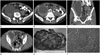
His chest X-ray showed several calcified nodules and fibrosis in the right upper lung field, indicating old healed TB, but it showed no interval changes when compared with the previous X-ray image which was taken 6 years ago. An abdominal computed tomography (CT) scan detected an oval-shaped cystic mass (8.3 × 6.5 × 20.9 cm) in the right lower retroperitoneal space. The mass had a well-defined outer margin, an irregular inner margin, wall enhancement, and hemorrhage. This mass compressed the right psoas muscle but it was relatively well demarcated from the muscle (Fig. 1A, B). There were focal wall calcifications within the mass (Fig. 1C). Several necrotic lymph nodes were observed around the mass in the right obturator space, and there was no evidence of ascites (Fig. 1D). Based on the radiographic findings, we considered cystic tumors that occur in the retroperitoneal space such as complicated cystic lymphangioma, mucinous cystadenocarcinoma, neurogenic tumors with cystic degeneration, or other undifferentiated sarcomas. Total surgical excision of the mass and adjacent lymph nodes was performed (Fig. 1E). Histopathologic examination by hematoxylin and eosin staining revealed granulomatous inflammation and caseous necrosis consistent with TB abscess (Fig. 1F); however, acid-fast staining and polymerase chain reaction test for Mycobacterium tuberculosis were negative. Immunohistochemical staining for D2-40, CD34, CD31, and calretinin was negative. There was no endothelial lining along the inner wall of the lesion, which ruled out the possibility of a pre-existing mesothelial or lymphangitic cyst. Anti-TB therapy with isoniazid, rifampicin, pyrazinamide, and ethambutol was administered for 6 months. After the completion of treatment, the patient did not develop any further symptoms during the follow-up.
DISCUSSION
Retroperitoneal abscesses are a uncommon clinical problem and they have a high mortality rate. They can occur as complications of other diseases such as perforated appendicitis, diverticulitis, perforated colonic or duodenal cancer, Crohn's disease of the bowel, pancreatitis, or trauma. They are often caused by polymicrobial infections, and the common pathogens are Escherichia coli, Klebsiella pneumoniae, Enterococcus spp., and Staphylococcus aureus. However, abdominal TB abscesses are uncommon in immunocompetent patients. Pathogenesis of such cases can be through hematogenous or lymphatic dissemination from active pulmonary TB or a direct extension from an adjacent organ (4).
Extrapulmonary TB abscess can present with variable radiologic features. It is difficult to differentiate it from lymphoma, malignant tumors, or various inflammatory conditions. Furthermore, the differential diagnosis becomes more complex when extrapulmonary TB presents as a soft tissue mass, showing involvement of the peritoneum, or an abscess. Epstein and Mann (5) discussed that it is important to differentiate other neoplasms from TB abscess. Therefore, the mass in our case should also be differentiated from cystic tumors occurring in the retroperitoneum.
Retroperitoneal tumors with a cystic component can be cystic lymphangiomas, mucinous cystic tumors, or neurogenic tumors with cystic degeneration. Also, malignant tumors can resemble a cystic mass if they have extensive necrosis. Unlike in this case, typical cystic lymphangiomas and mucinous cystic tumors have a thin, smooth wall and may have septa within the lesion. It is known that among neurogenic tumors, schwannomas can display cystic degeneration as well as focal areas of punctate calcification. However, they can be differentiated from TB abscess because schwannomas are seldom accompanied by necrotic lymph nodes. Among malignant tumors, retroperitoneal leiomyosarcomas tend to develop massive cystic degeneration. They can show central necrosis more commonly than the other sarcomas, whereas calcifications are not typically present. Furthermore, sarcoma accompanied by lymphadenopathy is quite rare (6, 7, 8).
Chuang et al. (9) reported a case of vertebral TB presenting as a large retroperitoneal cyst which has radiographic similarities with our case. There are also several reports of an iliopsoas abscess that originated from spinal TB (10). If there were findings suspicious for spinal TB or active pulmonary TB, TB abscess would have been highly suggested on CT imaging in our case. However, there was no evidence of vertebral TB or active pulmonary TB in our patient.
In conclusion, this is a rare case of TB abscess presenting as a large retroperitoneal cystic mass without active pulmonary or vertebral involvement. The diagnosis was based on CT imaging and histopathology. To the best of our knowledge, there are no published reports of retroperitoneal TB abscess which was confirmed by both CT scan and surgical pathology in the Korean literature. As TB remains endemic in Korea and with the continued increase in multidrug-resistant TB infections, it is important to know various imaging features of extrapulmonary TB including TB abscess. In this respect, this rare case provides an uncommon yet important differential diagnosis of a retroperitoneal cystic lesion.
 XML Download