

 PDF
PDF ePub
ePub Citation
 Print
Print


INTRODUCTION
Hemangioblastoma is a benign vascular neoplasm of the central nervous system. Although hemangioblastoma is the most common primary neoplasm in the adult cerebellum, it is a relatively rare vascular tumor of the spine, representing 1.6-5.8% of all spinal tumors (1). Spinal hemangioblastoma may occur sporadically or as a component of von Hippel-Lindau (VHL) syndrome. VHL syndrome is an autosomal dominant disorder, and it is caused by germline mutations of the VHL tumor supressor gene located on the distal part of the short arm of the third chromosome (3p25-26) (2). The prevalence of VHL syndrome ranges from 1:40000 to 1:50000 (3). VHL syndrome manifests as central nervous system hemangioblastomas, renal cysts, and renal cell carcinomas. Other lesions include retinal angiomas, pheochromocytomas, pancreatic cysts, and epididymal cystadenomas (4). Patients with VHL syndrome-associated spinal hemangioblastomas tend to present with neurological symptoms and signs at a younger age, and have multiple small lesions (5, 6, 7). When VHL syndrome-associated spinal hemangioblastomas are represented by multiple, tiny to small lesions, they can mimic other spinal tumors or disease including metastasis. It is important to know the clinical and imaging characteristics of VHL syndrome-associated spinal hemangioblastomas for appropriate image interpretation and management of the patient. The author reports a relatively rare case of VHL syndrome-associated spinal hemangioblastomas represented by multiple small lesions, with an emphasis on the magnetic resonance imaging (MRI) findings and discussion of the clinical characteristics of VHL syndrome-associated spinal hemangioblastomas.
CASE REPORT
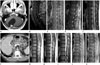
A 32-year-old man presented with gait disturbance and dizziness since 6 weeks. He had received laser therapy for retinal capillary angioma in his right eye, 8 years before he was referred to our hospital. After that, he was blind in the right eye. His father had died of an unknown cervical spinal cord lesion. On physical examination, he was not able to walk correctly in tandem gait, but, he was not swaying or tilting in ordinary gait. He had a wide-based gait. His sensory and motor functions were intact. These physical examination findings suggested a cerebellar lesion. Brain MRI demonstrated a 5 cm-sized, well-defined, thin and non-enhancing walled, intra-axial cystic mass with an intensely enhancing mural nodule in the right cerebellar hemisphere (Fig. 1A). For further evaluation, whole spinal MRI and abdominal computed tomography (CT) were performed. Spinal MRI showed multiple, tiny to small, intramedullary lesions, located in the superficial dorsal aspects of cervical, thoracic, and lower spinal cord including cauda equina. The lesions showed homogeneous, intense contrast-enhancement after gadolinium administration, and iso to low signal intensity on both T1-weighted image (T1WI) and T2-weighted image (T2WI) (Fig. 1B-E). Among the spinal lesions, the largest lesion measuring 8 mm was associated with peritumoral edema but no syrinx at the middle thoracic level (Fig. 1E). Although the vascular flow voids were absent, wavy contrast-enhancing prominent vessels were seen adjacent to the relatively larger spinal lesions (Fig. 1C). Abdominal CT demonstrated multiple pancreatic cystic masses with small septal calcifications (Fig. 1F), multiple renal cortical cysts, and a hepatic cyst. The clinical and imaging findings were suggestive of VHL syndrome. The genetic testing for VHL syndrome was positive. He underwent excisional surgery for the cerebellar mass. Pathologically, the mass was proved to be a hemangioblastoma. At 4 months postoperatively, his gait returned to almost normal. After one year, follow-up spinal MRI showed a newly developed small syrinx adjacent to the largest spinal lesion observed previously and more prominent peritumoral edema, while there were no other changes in the remaining spinal lesions (Fig. 1G-K). In view of the clinical and imaging findings, the spinal lesions were diagnosed as spinal hemangioblastomas.
DISCUSSION
Spinal hemangioblastomas are relatively rare vascular tumors of the spine, representing 1.6-5.8% of all spinal tumors (1). Spinal hemangioblastomas may occur as a sporadic isolated lesion or as a component of VHL syndrome. It has been reported that spinal hemangioblastomas are more commonly found in patients with sporadic lesions than in patients with VHL syndrome (1, 5, 6). However, more recent studies have shown different results (7, 8). Conway et al. (8) found that the incidences of spinal hemangioblastomas were 47% in patients with VHL syndrome and 12% in patients with sporadic disease. According to Takai et al. (7), spinal lesions were much more prevalent in patients with VHL syndrome (88.2%) than in patients with sporadic disease (20.5%). These results are based on the availability of high resolution imaging with high detectability of spinal lesions (7). Mean age at symptom onset is lesser in patients with VHL syndrome than in patients with sporadic spinal lesion, because most of the patients with VHL syndrome tend to have neurological symptoms at an earlier age due to cerebellar lesions. In patients with VHL syndrome, spinal hemangioblastomas are often multiple and small (1, 5, 6, 7). The small spinal lesions are usually asymptomatic. Symptoms of relatively large spinal hemangioblastomas are similar to those of other spinal canal tumors and include sensory change, motor disturbance, and pain (1). Because small spinal hemangioblastomas in VHL syndrome are usually asymptomatic, symptoms can help to differentiate mutiple small spinal hemangioblastomas from other diseases that may produce small, enhancing foci, such as metastatic tumors, sarcoidosis, tuberculosis, neurosyphilis, fungal infection, and multiple sclerosis, which may be symptomatic even when their enhancing lesions are small (5). Patients with VHL syndrome tend to have a high risk of recurrence and development of new lesions. Incomplete excision is the most common cause of recurrence (1, 5, 6, 7). The lower spinal cord including the conus medullaris and cauda equina is more frequently involved in patients with VHL syndrome, whereas sporadic spinal hemangioblastomas most often occur in the cervical and thoracic regions, and lower spinal lesions are rare (7). On MRI, spinal hemangioblastomas are usually hypointense to isointense on T1WI and isointense to hyperintense on T2WI, when compared with signal intensity of the spinal cord. Gadolinium-enhanced T1WI usually shows uniform intense enhancement of the mass. Small lesions are often isointense and thus difficult to differentiate from the spinal cord. Therefore, gadolinium-enhanced T1WI is important for the evaluation of lesions suggestive of small hemangioblastomas particularly in VHL syndrome (6). Chu et al. (5) described several characteristic MRI findings of spinal hemangioblastoma including a well-demarcated margin, intense contrast enhancement, superficial location of the intramedullary tumors reflecting the subpial location of the intramedullary lesions, most often in the posterior aspect of the spinal cord, the relatively large size of the syrinx as compared with the size of the intramedullary tumor, and the presence of vascular flow voids in or around medium-sized to large tumors. Spinal hemangioblastomas can also be found either in an intradural extramedullary or an extradural location (1, 5). The frequency of syrinx formation is high in cases of intramedullary tumors including ependymoma, astrocytoma, and hemangioblastoma. About 50-70% of spinal hemangioblastomas have been found to be associated with a syrinx (1, 5, 6). The pathogenesis of syrinx formation in spinal hemangioblastoma remains unclear (5). Transudation from the tumor vessels and secretion by tumor cells are generally believed to be the major causes of syringeal development in patients with intramedullary tumors (9). In our case, a small syrinx developed in the area of peritumoral edema adjacent to the largest spinal lesion during the one-year follow-up study (Fig. 1H, I, K). It is likely that peritumoral edema results from local venous congestion caused by a hypervascular tumor such as spinal hemangioblastoma, which has an arteriovenous shunt, and from the production of an edema-promoting factor by the neoplasm (10). In the present case, only one lesion that was the largest lesion was associated with peritumoral edema, which was more prominent in the one-year follow-up MRI (Fig. 1F, I).
In conclusion, patients with multiple small spinal hemangioblastomas involving the lower spinal cord, conus medullaris, and cauda equina, are more likely to have VHL syndrome. Small spinal lesions are usually asymptomatic. The author reports a case of VHL syndrome-associated spinal hemangioblastomas represented by multiple, tiny to small spinal lesions, with an emphasis on the clinical and imaging characteristics of VHL syndrome-associated spinal hemangioblastomas.
 XML Download