

 PDF
PDF ePub
ePub Citation
 Print
Print


INTRODUCTION
Gastric cancer is the fourth most common cancer worldwide (1) with a particularly high incidence in Asia (2). To reduce mortality from gastric cancer, it is essential to choose an optimal therapeutic approach, which depends on early detection and accurate preoperative staging.
Computed tomography (CT) has been the modality of choice for preoperative evaluation and staging patients with gastric cancer. Multidetector row CT (MDCT) with thin collimation offers near-isotropic imaging of the stomach and provides high quality multiplanar reformation. Dynamic contrast material-enhanced CT offers superior differentiation of tumor tissue from normal mucosa with adequate distention of the stomach using water as a negative contrast agent (3). However, CT plays only a limited role detecting lesions in patients with early gastric cancer (EGC) (1, 4).
18F-fluorodeoxyglucose positron emission tomography/computed tomography (18F-FDG PET/CT) is a useful diagnostic technique in clinical oncology. 18F-FDG PET/CT is highly accurate for determining resectability and detecting distant metastatic disease at the time of initial diagnosis. Various levels of FDG uptake have been found during primary tumor detection. Mucinous carcinoma and signet ring-cell carcinoma tend to show significantly lower FDG uptake than that of other histologic types of gastric cancer (5). The success rate of detecting EGC using 18F-FDG PET is < 50%, but detection rates of 62-98% have been reported, depending on the histological characteristics of the advanced gastric cancer (AGC) (6). However, no studies have addressed the use of combined hydro-stomach CT (S-CT) and 18F-FDG PET/CT to detect gastric cancer.
Therefore, the aim of this study was to retrospectively compare the diagnostic accuracy of S-CT with 18F-FDG PET/CT for detecting primary gastric cancer and to determine whether the combination of the two techniques improves diagnostic performance.
MATERIALS AND METHODS
Patients
This retrospective study was approved by our Institutional Review Board, and informed consent was waived. A total of 329 patients were pathologically confirmed with gastric cancer in our hospital from December 2009 to November 2012 and underwent S-CT.
Of the 329 patients, 76 were excluded from analysis for one of the following reasons: 1) only underwent S-CT without 18F-FDG PET/CT scanning (n = 20); 2) post-endoscopic submucosal dissection (ESD) status (n = 43); and 3) post-endoscopic clipping status or improper gastric distention (n = 13). After excluding these 76 patients, the final study group comprised 253 patients (age range, 35-86 years; mean age, 64.6 years; 176 men, age range, 35-86 years; mean age, 64.5 years and 77 women, age range, 38-86 years; mean age, 64.6 years).
Proof of Tumor Burden
Pathological proof of all lesions was obtained after ESD or surgery, which included ESD (n = 45), subtotal gastrectomy (n = 174), or total gastrectomy (n = 34). All 253 patients had a total of 267 confirmed adenocarcinomas (well-differentiated, 37; well- to moderately-differentiated, 5; moderately differentiated, 139; moderate to poorly differentiated, 19; poorly differentiated, 67). The specimens were histopathologically analyzed for depth of gastric wall invasion. Of the 267 lesions, 116 were classified as T1a, 63 as T1b, 26 as T2, 33 as T3, and 29 as T4a, according to the pathological TNM staging system developed by the 7th American Joint Committee on Cancer and the International Union Against Cancer (7).
CT Protocol
S-CT images were obtained from two scanners: a 64-channel CT scanner (Aquilion 64; Toshiba Medical Systems Co., Tokyo, Japan) in 232 patients, and a 128-channel CT scanner (Somatom Definition Flash; Siemens Medical Systems, Forchheim, Germany) in 21 patients. The following scanning parameters were used for the 64-MDCT scanner: collimation, 64 × 0.5 mm; pitch, 0.828; and rotation time, 0.6 second; and for the 128-MDCT: collimation, 128 × 0.625 mm; pitch, 0.8; rotation time, 0.5 second. The kilovoltage (kV) and effective tube current-time charge (mAs) were 120 kV and 200-250 mAs, respectively.
In our study, 500-1000 mL tap water was administered as an oral contrast medium to each patient immediately before CT to distend the stomach. Each patient had fasted for > 6 hours, and had received a 2 mL/kg intravenous dose (total volume, < 150 mL; 3 mL/sec) of nonionic contrast material (Ultravist 300; Schering, Berlin, Germany) through an 18-G angiographic catheter inserted in a forearm vein using an automatic power injector (Stellant D; Medrad, Pittsburgh, PA, USA). After obtaining unenhanced CT images, portal venous phase images were acquired 60-70 seconds after administration of the contrast medium. S-CT scans were obtained in the prone position, and the scanning field ranged from the diaphragmatic dome to the anal verge. All examinations were performed during deep inspiration. Axial S-CT images were reconstructed with 5 mm section thickness and a 5 mm reconstruction interval for clinical interpretation, in addition to axial images. Coronal multiplanar reconstruction (MPR) images were reconstructed with a 3 mm section thickness at a 3 mm interval.
18F-FDG PET/CT Protocol
All patients were instructed to fast for 8 hours (except for glucose-free oral hydration) before the PET examination, and blood glucose concentration was measured and confirmed to be 140 mg/dL. Intravenous injections of 5.5 MBq of 18F-FDG/kg body weight were administered. All patients were kept lying comfortably during the 60-minute uptake period and voided urine before being positioned supine on the scanning table. Integrated FDG PET/CT scanning was performed using a combined PET/CT scanner (Philips Gemini, 16, Best, the Netherlands). The first unenhanced CT scan with a 16 slice scanner was performed from the ear to the mid-thigh 60 minutes after the 18F-FDG injection using the following parameters: 120 kVp; 250 mA; rotation time, 0.5 second; helical thickness, 5 mm; 24 mm per rotation (speed); and a 128 × 128 matrix. A PET scan was then acquired from the level of the ear through the mid-thigh in three-dimensional mode at 1 minute per bed position. The PET unit had an axial field of view of 18 cm and a spatial resolution of 4.5 mm in full width of half maximum at 1 cm from the center. PET data were reconstructed iteratively using the row action maximum likelihood algorithm. The reconstructed CT, PET, and fused PET/CT images were displayed in axial and coronal planes. The median interval between S-CT and 18F-FDG PET/CT was 1 day (range, 1-22 days).
Image Analysis
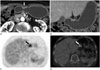
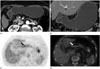
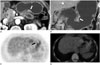
Two radiologists (with 11 and 4 years experience in radiology, respectively) who were familiar with interpreting S-CT and whole body PET/CT examinations, reviewed the S-CT and 18F-FDG PET/CT scans. The radiologists knew that the patients had been referred for a gastric cancer evaluation but were unaware of all other information regarding the patient's detailed medical history, laboratory results, findings from other imaging modalities, and the final diagnosis. They independently reviewed the three S-CT and 18F-FDG PET/CT sets in a random order; 1) the S-CT set, 2) the 18F-FDG PET/CT set, 3) the combined set, i.e., the S-CT set and 18F-FDG PET/CT set. Each reading session was separated by 4 weeks to minimize recall bias. The images from each set were presented to each reader in a random order during each session. Differences in their assessments were resolved by consensus. If at least one of the two readers correctly indicated a gastric cancer lesion, it was regarded as a visible tumor. If both reviewers missed a gastric cancer lesion, it was regarded as an invisible tumor.
The stomach was divided into three segments along the longitudinal axis (from the gastroesophageal junction to the pyloric canal) of upper, middle, and lower thirds (8). If the gastric cancer was located across segments, the position of the gastric cancer was set where the lesion had the greatest involvement.
Both reviewers graded the likelihood for the presence of primary gastric cancer in 759 gastric segments based on a 4-point scale as follows: 1, definitely absent; 2, probably absent; 3, probably present; and 4, definitely present. The readers were aware that only scores of 3 and 4 would be considered gastric cancer for statistical analysis purposes. The presence of EGC was defined as mucosal enhancement with or without focal thickening in the gastric inner and/or middle layer during analysis of the preoperative S-CT scans for detecting primary tumors. Strong and focal FDG uptake combined with a delayed image was indicative of a malignant lesion during analysis of the preoperative 18F-FDG PET/CT scans for detecting primary tumors, but diffuse or segmental patterns without focally increased accumulation were interpreted as physiological uptake. Strong and focal FDG uptake lesions, which were invisible on S-CT, were considered malignant lesions. All images were reviewed using a local picture archiving and communication system (GE Medical Systems, Milwaukee, WI, USA).
Statistical Analysis
All lesions, EGCs, and AGCs were analyzed separately. An alternative-free response receiver operating characteristic (ROC) curve was fitted to each reader's confidence scoring based on the retrospective interpretation. The diagnostic accuracies of the S-CT and the combined sets were determined by calculating the area under each reader-specific ROC curve (Az) (9). Sensitivity, specificity, positive predictive value (PPV), and negative predictive value (NPV) were calculated for detecting gastric cancer in each of the modalities. Interobserver agreements for the confidence ratings to detect gastric cancer were analyzed with kappa statistics: < 0.40 = poor agreement; 0.41-0.74 = moderate/good agreement; and > 0.75 = excellent agreement. The McNemar test, logistic regression with the generalized estimating equation method, and the weighted least square method for repeated categorical data analysis were used to assess the statistical significance of any difference among the modalities (S-CT set, 18F-FDG PET/CT set, and combined set). All statistical computations were performed using MedCalc Software, v.12.7.8 statistical software (Mariakerke, Belgium), SAS 9.2 (SAS Institute, Cary, NC, USA), or SPSS 19.0 software (SPSS Inc., Chicago, IL, USA). A p-value < 0.05 was considered significant.
RESULTS
Of the 267 primary gastric cancers, 22 lesions were located in the upper segment, 91 in the middle segment, and 154 in the lower segment.
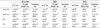
Each reader noted significantly higher Az values for diagnosing all primary gastric cancers and EGCs with the combined set than those for the S-CT set. The reader's Az values were not different between the combined set and the S-CT set for detecting AGCs (Table 1, Fig. 1).
In the consensus reading, sensitivities for detecting all primary gastric cancers and EGCs were significantly higher with the combined set (66.3% for all lesions; 50.8% for EGCs) than those with the S-CT set (61.4% for all lesions; 40.2% for EGCs) and with the 18F-FDG PET/CT set (42.3% for all lesions; 27.9% for EGCs) (Fig. 2). The combined set also had significantly higher sensitivity (98.9%) than that of the 18F-FDG PET/CT set (72.7%), but no significant difference was observed with the S-CT set (97.7%) for detecting AGCs (Fig. 3). Specificities were significantly higher with the 18F-FDG PET/CT set (100% for all lesions and EGCs) than those with the S-CT set (98.8% for all lesions; 98.2% for EGCs) and the combined set (98.8% for all lesions; 98.2% for EGCs) for detecting all primary gastric cancers and EGCs. The PPVs for detecting all primary gastric cancers and EGCs were also significantly higher with the 18F-FDG PET/CT set (100% for all lesions and EGCs) than those with the S-CT set (96.5% for all lesions; 92.3% for EGCs) and with the combined set (96.7% for all lesions; 93.8% for EGCs). In addition, the NPVs for detecting all primary gastric cancers and EGCs were significantly higher with the combined set (84.4% for all lesions; 78.7% for EGCs) than those with the S-CT set (82.5% for all lesions; 75.2% for EGCs) or the 18F-FDG PET/CT set (76.2% for all lesions; 72.0% for EGCs) (Table 2).
Six false-positive lesions were noted by both readers in the S-CT set during the consensus reading. All six lesions showed mild mucosal enhancement or subtle elevated lesions on portal phase S-CT scans. These lesions were not detected on the 18F-FDG PET/CT set. Endoscopic or pathological findings of these lesions suggested gastritis or non-specific lesions. However, these lesions were regarded as negative lesions on the combined set.
Interobserver agreement for detecting all lesions, EGCs, and AGCs was excellent (k = 0.764-0.914) (Table 3).
DISCUSSION
Gastric cancer is one of the most common malignant tumors of the alimentary tract. Improved early diagnosis, accurate clinical staging, and optimal surgical procedures are essential to improve prognosis (10).
CT has been the modality of choice for the preoperative evaluation and staging patients with gastric cancer. However, its use for detecting gastric cancer is limited because CT has a primary tumor detection rate of 85-95% in patients with AGC (1, 11, 12) and plays only a limited role detecting lesions in patients with EGC. Studies in which dynamic or multiphase scanning was used to evaluate EGC achieved detection rates of 44-93.5% (1, 3, 13, 14, 15, 16). In our study, sensitivities of 40.2% and 97.7% were observed in patients with EGC and AGC, respectively, using S-CT alone. S-CT alone was limited to detect lesions in patients with EGC.
18F-FDG PET has no role in primary gastric cancer detection due to its low sensitivity, particularly in EGC (17). Our study also showed sensitivity of 27.9% in patients with EGC using 18F-FDG PET/CT alone. Newly developed PET-CT technology using computer software fuses the PET metabolic-change image with a three-dimensional image of the corresponding anatomical location on the CT image, which improves diagnostic accuracy of cancer metastasis to the lymph nodes and organs distant from the tumor (10). However, the stomach was difficult to evaluate on 18F-FDG PET/CT because the 18F-FDG PET/CT protocol did not include expanding the stomach and used non-contrast CT. 18F-FDG PET/CT and gastric distension using a mixture of milk and Diatrizoate Meglumine results in more obvious contrast between the normal stomach wall and the lesion, but it does not significantly improve diagnostic accuracy (17). Zhu et al. (18) reported that gastric cancer approximately 1.2 cm in diameter is detectable using a mixture of milk and Diatrizoate Meglumine for gastric distension on PET/CT imaging. Kamimura et al. (19) reported that the accuracy of cancer diagnosis increases by having the patient drink water to increase gastric volume prior to 18F-FDG PET. However, any potential improvement in PET/CT diagnostic accuracy for gastric cancer using negative oral contrast agents compared to positive oral contrast agents needs to be further evaluated.
In this study, 253 patients were subjected to a qualitative diagnosis of primary gastric cancer using a combination of S-CT and 18F-FDG PET/CT. Overall sensitivities showed that the combined set of S-CT and 18F-FDG PET/CT provided significantly higher sensitivities for detecting all primary gastric cancers and EGCs than those of S-CT and 18F-FDG PET/CT alone.
Because EGC can only be detected by either S-CT or 18F-FDG PET/CT, the combined set resulted in a significantly higher detection rate than that of S-CT or 18F-FDG PET/CT alone. For example, linitis platisca of the stomach, in which gastric endoscopy is normal, can be detected by 18F-FDG PET/CT (20). In addition, the combined set also had a significantly higher NPV for detecting all primary gastric cancers and EGCs compared to that of S-CT and 18F-FDG PET/CT alone but was not different for detecting AGCs.
The combined S-CT and 18F-FDG PET/CT set had the following advantages. First, gastric cancer lesions often obscured by physiological uptake on 18F-FDG PET/CT can be identified on S-CT, which provides detailed anatomical information. Second, if EGCs, which have a low detection rate on S-CT, are identified on 18F-FDG PET/CT, it is possible to review the S-CT backwards to find the primary cancer. Although sensitivities for detecting all primary gastric cancers and EGCs with the combined set were 66.3% and 50.8% respectively, it was valuable to add 18F-FDG PET/CT to S-CT to detect and localize primary gastric cancer.
This study had some limitations. First, this study was a retrospective, single institution study over a defined period. Second, we did not include CT gastrography, which is helpful for detecting primary gastric cancer (3). Therefore, a further comparative study is needed using S-CT and CT gastrography to detect primary gastric cancer. Third, this study was conducted in a highly selected patient population with primary gastric cancer diagnosed by endoscopic biopsy. Thus, it was unclear to what degree the current findings can be generalized to a wider population. Fourth, arterial phase images were not obtained. We obtained images in the portal venous phase (70 seconds). Lee et al. (16) reported that helical CT with a two-phase scan including the mucosal phase is efficient for identifying EGC enhancement patterns. However, it is controversial whether there is added value of arterial phase imaging to reveal gastric cancer on CT. Fifth, axial S-CT images were reconstructed with 5-mm section thickness and a reconstruction interval. Choi et al. (21) reported that axial CT images used for preoperative gastric cancer staging should be reconstructed using 3-mm section thickness and a 2-3 mm reconstruction interval. Coronal and sagittal MPR images are also reconstructed with a 3-mm section thickness and interval. However, other reports used a 5-mm section thickness for axial CT images (3, 8), whereas we included coronal MPR images with a 3-mm section thickness for detecting gastric cancer. Sixth, 18F-FDG PET/CT was performed without gastric distension. 18F-FDG PET/CT with gastric distension using a negative or positive oral contrast agent increases the detection rate of primary gastric cancer.
In conclusion, detecting primary gastric cancer for localization and staging is very important, and our results show that the combined S-CT and 18F-FDG PET/CT set provided a significantly higher detection rate for primary gastric cancer than that of S-CT or 18F-FDG PET/CT alone, particularly for EGCs. Higher diagnostic accuracies were obtained with a combined set than that with S-CT alone. Therefore, combined reading of S-CT and 18F-FDG PET/CT is recommended to ensure better detection during preoperative evaluations for primary gastric cancer.


 XML Download