

 PDF
PDF ePub
ePub Citation
 Print
Print


INTRODUCTION
A fibrous pseudotumor is a benign fibroproliferative tumor arising from the paratesticular tissues. The majority of the reported cases show an involvement of the tunica vaginalis and the others are associated with the epididymis, spermatic cord and other surrounding connective tissues (1, 2, 3). To the best of our knowledge, there are only rarely reports on the experiences in diagnosing with both ultrasonography (US) and magnetic resonance imaging (MRI), even more rarely are reports on diagnosing with dynamic contrast-enhanced MRI. In this article, we present a case of fibrous pseudotumor of the tunica vaginalis presenting as painless palpable masses on the right scrotum in a 33-year-old man, with US and MRI findings including the dynamic study.
CASE REPORT
A 33-year-old man presented to our institution with painless palpable masses on the right scrotum for 2 years. The patient did not have any history of trauma or infection. Laboratory studies were unremarkable. On physical examination, the right scrotum was swollen and a small non-tender hard mass was palpated.
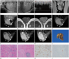
An US with 5- to 12-MHz linear array transducer (iU22, Philips Healthcare, Eindhoven, The Netherlands) revealed multiple well-demarcated, round to oval, iso- to slightly hypoechoic nodular lesions along the tunica vaginalis in the right scrotal sac (Fig. 1A, B). The largest one measured about 1.5 cm in size. Some nodules showed posterior shadowing without any calcification. On Doppler study, multifocal small areas of color flow signal were noted (Fig. 1C). A small amount of fluid collection was also seen in the right tunical sac. Both testes and epididymides were normally well seen.
On MRI (Intera, 1.5-T, Philips Medical Systems, Best, The Netherlands), multiple sharply demarcated, round to oval nodules were seen in the right scrotal sac. The lesions showed a markedly low signal intensity (SI) on T2-weighted image (T2WI), a slightly low SI on T1-weighted image (T1WI) and a mild enhancement on the contrast-enhanced T1WI (Fig. 1D-G). A dynamic contrast-enhanced study with sagittal fat suppression T1WI was performed. Four post-contrast images were obtained at 30, 90, 180, and 300 seconds after the administration of the contrast agent. The lesions showed a mild and progressive enhancement persisting through the delayed phase (Fig. 1H-K). The provisional diagnosis was a fibrous pseudotumor of the testicular tunic and the differential diagnosis included a mesothelioma.
The patient underwent a surgical excision and the intraoperative findings revealed up to 1 cm sized multiple irregular shaped hard nodules attached to the testis and epididymis without direct invasion. A total excision of the mass was performed with some part of the tunica vaginalis preserving testis and epididymis. Pathologically, the gross specimen showed the lesion in the right scrotal tunica vaginalis measuring 5.7 × 2.7 × 2.9 cm in size. Multiple firm consistent nodules were attached to the surface of the tunica vaginalis (Fig. 1L). Microscopically, the lesion showed abundant sclerotic and fibrous tissue surrounding vascular structures and largely hypocellular tissue with clusters of inflammatory cells (Fig. 1M, N). An immunohistochemical study was performed and the tumor cells were unusually not reactive for smooth muscle actin, desmin, CD34, and S-100 protein (Fig. 1O, P). An additional stain for immunoglobulin G4 (IgG4), done to rule out an IgG-related disease was also negative. Finally, the lesion was pathologically confirmed as fibrous pseudotumor.
DISCUSSION
Paratesticular tumors originate in the intrascrotal surrounding structures such as testicular tunic, epididymis or spermatic cord (2, 3, 4). First reported by Balloch in 1904, a fibrous pseudotumor is a benign reactive process of fibrous proliferation in the paratesticular tissue (1, 2, 3, 4). It has been known by several synonyms including inflammatory pseudotumor, pseudofibromatous periorchitis, chronic proliferative periorchitis, fibrous mesothelioma, and reactive periorchitis (1, 4). It is most likely that the etiology of this entity is closely related to an inflammatory process such as epididymitis, infected hydrocele, previous surgery or trauma (1). Although a fibrous pseudotumor is rare and comprises about 6% of paratesticular masses, it is the second most common mass involving paratesticular tissues, second only to the adenomatoid tumor of the epididymis (1, 2). It has a peak incidence in the third decade of life, but can occur at any age (4, 5). Generally, the size of the tumor ranges from 0.5 to 8 cm in maximal diameter, with the largest one reported to be 25 cm (4). While intratesticular tumors are more likely to arise from the right side, paratesticular tumors have no reported predilection of laterality (1, 2).
The patients usually present with a palpated single or multiple, painless, firm scrotal nodules. Less commonly, the lesion may also appear as a diffuse thickening of the testicular capsule (3, 5). Approximately 30% are related to a history of prior trauma or infection, supporting the reactive pathogenesis of the lesion (2, 3, 5). Nearly 50% of the cases represent a hydrocele or hematocele as the most frequently associated findings (2, 3, 4, 5).
On microscopic examination, the fibrous pseudotumor shows proliferative fibroblasts and other inflammatory cells intermixed with collagen bundles in the dense hyalinized fibrous tissue (1, 2, 5). Small capillary vessels or calcifications may also be present within the lesion. Immunohistochemical staining is usually positive for smooth muscle-specific actin, vimentin, common muscle actin and negative for keratin, desmin and S-100 protein (4).
An US examination with a high-frequency linear probe is the first modality of choice for the detection and evaluation of the scrotal lesion. On gray-scale US, fibrous pseudotumors appear as single or multiple, hypoechoic or hyperechoic nodules, depending on the amount of collagen, fibroblasts or calcifications (2, 3, 5, 6). The lesion may show posterior shadowing without calcification due to the dense fibrous and collagenous content. On color Doppler US, a small to moderate amount of vascularity can be seen within the lesion (5). Our case appeared as multiple isoechoic nodular lesions in the right scrotum with minimal vascularity and some nodules showed posterior shadowing without any calcification.
A MRI may help to provide a better tissue characterization and thus a better depiction of the fibrous nature of the mass (7). It also provides additional information of adjacent tissues with a wider field of view. The mass usually shows homogeneous low SI relative to the testis on both T1- and T2WIs due to dense fibrotic tissue (2, 3, 6, 7). Typically, the mass shows a minimal enhancement on the contrast-enhanced image, but the enhancement can be variable depending on the capillary network in the lesion (5, 7). In our case, the mass showed dark SI on T2WI, slightly low SI on T1WI and a mild enhancement on the contrast-enhanced T1WI. Although reported cases including dynamic study are limited, the lesion is generally thought to show slow but persistent enhancement due to its fibrous nature as presented in our case (7).
The radiologic differential diagnosis includes adenomatoid tumor, leiomyoma, lipoma, and mesothelioma. An adenomatoid tumor is a benign tumor of mesothelial origin and the most common tumor of the epididymis, accounting for 30% of all paratesticular tumors. The US shows a well-defined iso- or hypoechoic ovoid lesion with or without a cystic portion (8). The MRI shows an intermediate SI on T1WI, a slightly low SI on T2WI and a variable enhancement (7). The leiomyoma is the second most common neoplasm of the epididymis. On US, multiple recurrent narrow areas of shadow suggesting transition zones and whorl-shaped echo pattern are characteristic findings (9). The lipoma tends to be a well-defined homogeneous hyperechoic mass without internal vascularity on US, but may be rarely hypoechoic or heterogeneous in echotexture. A characteristic of fat tissue is a homogeneous high SI without enhancement on both T1 and T2WIs on MRI. The fat suppression technique or the in- and out-of-phase imaging may also help defining the fat component within the tumor (7). As the tunica vaginalis is a layer of reflected peritoneum, a malignant mesothelioma can also occur, especially in individuals with a history of asbestos exposure. Suggestive US findings are multiple heterogeneous or hyperechoic nodules within the enlarging hydrocele space and a hyperemia of the involved tunica vaginalis (10). On MRI, the lesion is iso- to hyperintense on T1WI, hyperintense on T2WI and it is well enhanced. Although fibrous pseudotumor can occur as both single and multiple lesions, the multiplicity of the lesion helps to narrow the differential diagnosis into fibrous pseudotumor and malignant mesothelioma (7, 8, 9, 10).
The treatment of the fibrous pseudotumor includes the excision of the mass and involved testicular tunic, sparing the testis (1, 2, 3, 6). However, a radical orchiectomy has been frequently performed in many cases despite the benign nature because of the rarity of this tumor. US and MR imaging facilitate an early recognition of the intactness of testis and the establishment of an appropriate treatment plan (2, 7). An orchiectomy is recommended in some rare cases of diffuse band-like fibrous tissue surrounding the entire testis (4).
In summary, we report a case of fibrous pseudotumor arising from the tunica vaginalis of the right scrotum. Characteristic imaging findings on US and MRI are helpful in the detection and diagnosis of the fibrous pseudotumor, thus avoiding an unnecessary treatment such as a radical orchiectomy.
 XML Download