

 PDF
PDF ePub
ePub Citation
 Print
Print


INTRODUCTION
The complex anatomy of the great vessels in the mediastinum give rise the occasional occurrence of anomalies. These anomalies are usually associated with congenital heart disease. An anomalous left brachiocephalic vein (ALBCV) is rare. The incidence of ALBCV with congenital heart disease is 0.2-1%, whereas in the general population, the incidence of this anomalous vein without congenital heart disease is 0.06-0.37% (1, 2). Most reports on ALBCV are on aberrant left BCV, and there are few reports on double left BCV (3). The double left BCV, also designated as the double left innominate vein or a duplication of the left innominate vein, is an exceptionally rare congenital anomaly. The most common variant of this anomaly is the coexistence of a retroaortic left brachiocephalic vein and a normally placed left brachiocephalic vein (3).
Persistent left superior vena cava (PLSVC), while uncommon, is the most common thoracic venous anomaly. PLSVC occurs in 0.3% of the general population, but is more common in patients with congenital heart disease (4-4.3%) (4). Aberrant left BCV with PLSVC is very rare and was reported by Chen et al. (2). To our knowledge, double left BCV with PLSVC has not been reported yet. Here, we report a case of double left BCV with PLSVC in a 72-year-old male patient with no existing cardiac abnormality.
CASE REPORT
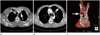
A 72-year-old male was admitted to our hospital due to an incidental lung mass on his chest radiograph. A chest CT scan was performed for further evaluation of the lung nodule, and revealed double left BCV and PLSVC (Fig. 1). The left BCV was divided into two branches: the anterior branch was a normally placed left brachiocephalic vein, and the posterior branch coursed below the aortic arch and drained into the right superior vena cava (SVC). The PLSVC was formed by the left internal jugular vein and the left subclavian vein.
The PLSVC connected to the right atrium via the coronary sinus. The right BCV and the right SVC were normal and the man not diagnosed with any other cardiovascular anomaly. Percutaneous needle biopsy was performed on the right middle lung nodule, and the histopathologic finding was squamous cell carcinoma of the lung. For treatment, right middle lobectomy with mediastinal lymph node dissection was performed.
DISCUSSION
Normally, the union of the left subclavian vein and the left internal jugular vein forms the left BCV behind the sternoclavicular joint. The left BCV passes across the mediastinum and courses obliquely inferior and anterior to the ascending aorta or the brachiocephalic artery to the right upper chest (3). Rarely, this vein takes an anomalous course. While the exact cause of an ALBCV is unknown, the most accepted mechanism is the interruption of the upper anastomosis between the right and left anterior cardinal veins (1, 2).
There are several classifications of ALBCV. Takada et al. (3) classified the types of aberrant and double left BCV according to the course of the aberrant left BCV. Chen et al. (2) suggested several theories and patterns of aberrant and double left BCV. The double left BCV without PLSVC had also been reported by Takada et al. (3) and Subirana et al. (5), one in the normal position and the other crossing beneath the aortic arch.
The classification of the embryological anomalies of SVC has been presented by Nandy and Blair (6) and Starck (7). The most common subtype of PLSVC results in the presence of both left and right SVCs. A bridging brachiocephalic vein may or may not be present. A single PLSVC is much rarer. Variations have also been reported in the insertion of PLSVC. In 90% of cases, the PLSVC connects to the right atrium via the coronary sinus and has no hemodynamic consequence. Our patient has double SVC: the right one is normal whereas the left one, formed by the left internal jugular vein and the left subclavian vein, is thinner and drains into the right atrium via the coronary sinus.
ALBCV accompanied by PLSVC is extremely rare. Webb et al. (8) reported that PLSVC is associated with the absence of BCV. Chen et al. (2) reported that the presence of PLSVC was significantly less common in situs solitus patients who had ALBCV than in those who did not (p < 0.05 in the comparison of the tetralogy of Fallot, the double outlet of the right ventricles, the ventricular septal defect, and the atrial septal defect subgroups). To our knowledge, double left BCV with PLSVC has not been reported yet.
ALBCV and PLSVC are usually incidental findings in many cases. In a non-contrast-enhanced CT scan, they can be misinterpreted as enlarged lymph nodes or central pulmonary arteries. In patients with other cardiac anomalies, diagnosis can become difficult. Carefully tracing these vascular structures through sequential images is the key to their differentiation.
Clinically, knowledge of these anomalies is important and should be considered when inserting a central venous catheter or in cardiac surgeries. These anomalies may cause technical difficulties during pacemaker insertion or central venous line placement via the left arm approach. For patients undergoing cardiac surgery, the superior vena caval cannulation for a cardiopulmonary bypass must be done in a more caudal fashion than usual to avoid obstruction of the retroaortic branch of the left BCV.
 XML Download