

 PDF
PDF ePub
ePub Citation
 Print
Print


INTRODUCTION
Although a large proportion of all solid testicular masses are malignant, testicular adrenal rest tumor (TART) is a benign intratesticular tumor. The reported prevalence of TART in patients with congenital adrenal hyperplasia (CAH) varies widely, even up to 94% (1). The appearance of TARTs on both gray-scale and color Doppler ultrasound (US) has been reported (2, 3, 4). Transition of the gray-scale and color Doppler US features of TART according to hormonal control is unclear, and US imaging of irregular speculated hyperechoic mass has not been reported. We herein report three cases of usual and unusual sonographic findings and long-term follow-up images correlated with hormonal transitions.
CASE REPORT
Case 1
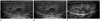
A 14-year-old boy with a neonatal diagnosis of CAH had been treated with prednisolone and fludrocortisone. CAH had been poorly controlled because a poor compliance. Laboratory studies revealed a level of adrenocorticotropic hormone (ACTH) and 17-hydroxyprogesterone (17-OHP) of 65.8 pg/mL (reference range, 10-60 pg/mL) and 41.93 ng/mL (reference range, 0.42-3.5 ng/mL), respectively. There was no reported history of testicular pain. Physical examination of the scrotum did not reveal palpable mass. US performed to assess testicular abnormalities demonstrated five well defined oval and round mixed hypoechoic masses around the mediastinum of the bilateral testes (two in the right, three in the left). The masses had no acoustic shadowing (Fig. 1A). Color Doppler US demonstrated nodular vascularity in most masses (Fig. 1B). The epididymides were unremarkable. On the basis of the elevated 17-OHP level and a history of CAH, US findings were compatible with TART and a biopsy was not performed. After improvement of the compliance and regularity of the treatment for 3 months, the levels of ACTH and 17-OHP became within the normal ranges. During a 7-year follow-up, the levels of ACTH and 17-OHP were well controlled. On US performed 4 years later, the dimensions of the lesions in both testes were decreased (right; 20 × 14 × 13 mm to 13 × 10 × 9 mm, left; 9 × 7 × 9 mm to 6 × 5 × 5 mm). The vascularity of the lesions was not changed. However, ill-defined hyperechoic lesions surrounding the previous masses had developed (Fig. 1C). On US performed 7 years later, the ill-defined hyperechoic lesions surrounding the masses were prominent though the dimensions and vascularity of the lesions in both testes were unchanged.
Case 2
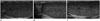
A 10-year-old boy with poorly controlled CAH diagnosed at 6-years-of-age presented with precocious puberty and short 328stature. Laboratory studies showed an ACTH level of 849.84 pg/mL and 17-OHP level of 103.39 ng/mL. His physical examination was unremarkable. He underwent scrotal US for investigation of testicular abnormalities, which demonstrated a small well defined oval hypoechoic mass (3 × 2 × 3 mm) in the right mediastinum testis (Fig. 2A). The mass had no acoustic shadowing. Color Doppler US demonstrated no vascular flow in the mass. On the basis of the elevated 17-OHP level and a history of CAH, US findings were compatible with TART and a biopsy was not performed. After improvement of the compliance and regularity of the treatment for 6 months, the levels of ACTH and 17-OHP became within normal range. During a 6-year follow-up, the levels of ACTH and 17-OHP were poorly controlled. Although a follow-up US 6 months later showed complete resolution of the lesion (Fig. 2B), on US reexamination 23 months later, three new well defined oval hypoechoic masses with nodular vascularity were evident in both mediastinum testes (Fig. 2C). On US performed 6 years later, the dimensions of the lesions in both testes were increased (right; 4 × 3 × 3 mm to 6 × 4 × 5 mm, left; 2 × 2 × 2 mm to 4 × 3 × 3 mm) though vascularity of the lesions in both testes were unchanged.
Case 3
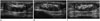
A 13-year-old boy with CAH diagnosed neonatally presented with precocious puberty and short stature. CAH had not been treated for 13 years. Laboratory studies showed an ACTH level of 1970 pg/mL and a 17-OHP level of 125 ng/mL. The patient underwent scrotal US, which demonstrated bilateral irregular speculated hyperechoic masses around the mediastinum of the testes without posterior acoustic shadowing and small oval well defined hypoechoic nodular lesions adjacent to the main mass in the left testis (Fig. 3A). Color Doppler US demonstrated central nodular vascularity in both testes (Fig. 3B). The right lesion measured 20 × 9 × 9 mm, and the left lesions measured 19 × 6 × 11 mm and 8 × 5 × 7 mm and 8 × 5 × 5 mm. Both testicular sizes were small; the dimension of the testis was 25 × 12 × 15 mm. Testicular tumor marker test results were negative. On the basis of the elevated 17-OHP level and a history of CAH, TART was suggested and a biopsy was not performed. After treatment for 3 months, the levels of ACTH and 17-OHP became within the normal range. During a 4-year follow-up, the levels of ACTH and 17-OHP were well controlled. A follow-up US 6 months later demonstrated no change in the size and shape of bilateral main masses, but complete resolution of the small masses adjacent to main mass in the left testis (Fig. 3C). This patient has been followed by US for the past 4 years, and his main testicular mass has been stable.
DISCUSSION
CAH is an inherited disorder of adrenal steroid synthesis that is caused by 21-hydroxylase deficiency, and which leads to glucocorticoid and mineralocorticoid deficiency and increase in ACTH secretion. It is thought that poor hormonal control, leading to high blood level of ACTH, is an important factor in the pathogenesis of TART inducing hyperplasia of adrenal-like cells within the testis (5). These tumors were first reported in 1940 (6). Since then several reports have described features of TART.
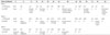
Morphology and size of TART are stable or changed depending on how CAH would be controlled. Hormonal control was assessed by serum measurements of ACTH and 17-OHP. Because ACTH has highly variable secretion and short half-life, subtle abnormalities in circadian ACTH secretion cannot be excluded for single measurement and 17-OHP is regulated by ACTH and indirectly reflects ACTH effect with longer half-life. Previous reports (7, 8) have suggested that TART may decrease in size with adequate hormonal control and alternatively may increase in size when hormone therapy is inadequate or when patients are noncompliant. Avila et al. (4) reported changes in size of TART were not related to the serum 17-OHP level in 11 patients. However, their reference level of 17-OHP as good control, < 1300 ng/dL was very high and they did not suggest exact specific cases serially in the study. Table 1 shows serial levels of 17-OHP and changes of TARTs in three patients. When patients were in well hormonal control, five hypoechoic masses decreased and then maintained stability in size and three masses disappeared, although two irregular hyperechoic masses were unchanged. When patient was in poor hormonal control, one and two masses in patient 2 reappeared and developed newly, then increased in size during follow-up. These findings suggested a correlation with TART size and hormonal control. During long follow-up, vascularity of the masses on Doppler US had not changed significantly regardless of hormonal control.
Sonographically, TART in most cases present as hypoechoic lesions peripherally located close to the mediastinum of the testis and are usually bilateral (9). Most masses in the three patients showed the same US findings except irregular speculated hyperechoic masses in patient 3. US imaging of irregular speculated hyperechoic masses bilaterally have not been reported. Previously reported hyperechoic masses were lesions larger than 2 cm, being hypoechoic with hyperechoic reflections (2) and lobular elongated homogeneous hyperechoic masses (3, 10). Although the masses in the three present patients were large and elongated, US findings of relatively homogeneous hyperechogenicity and speculated margin are unique. Pathologic evaluations of hyperechoic lesions have suggested that hyperechogenicity results from dense fibrotic stroma, fatty infiltration, and multiple interface between the stromal and tumor cell cluster (11). Accordingly, these bilateral masses remained stable in size and morphology during a 4-year follow-up, despite good hormonal control. Irregular speculated margin of the masses may be explained by proposed classification of TART (1). These masses in both atrophied testes of patient 3 may be in stage 5: irreversible damage of testicular parenchyma, peritubular fibrosis and peritubular hyalinization along seminiferous tubules due to no medication for 13 years before treatment. This classification may also explain a hypothesis that the ill-defined hyperechoic lesions surrounding the previous masses that developed in patient 1 during follow-up period are due to long-term consequence of fibrosis.
In summary, TART on US examination can change during a long-term follow-up period by increasing, decreasing, disappearing, or developing further, depending on hormonal control. Unique US finding of irregular speculated hyperechoic masses in the atrophied testes may be indicative of an advanced stage of disease.
 XML Download