

 PDF
PDF ePub
ePub Citation
 Print
Print


INTRODUCTION
Thoracic endovascular aortic repair (TEVAR) has been accepted as an alternative to open surgical repair in certain groups of patients (1). A potential complication caused by persistent or recurrent flow into the aneurysm sac is known as endoleak which can be classified to four types (2). Complex type II endoleaks occur due to persistent reverse blood flow through branch vessels (3). Common sites of type II endoleaks are the left subclavian artery and intercostal arteries with occasionally bronchial arteries (4). Arterial pressure in type II endoleaks is transmitted to the aneurysm sac, causing sac expansion. In this situation, patients remain at risk of aneurysm rupture (3). Treatment of type II endoleaks requires elimination of blood flow through branch vessels to relieve the aneurysm sac from systemic pressure (3). Various methods are introduced to manage type II endoleaks, such as the use of coils, plugs, or liquid embolic agents (histoacryl, thrombin, onyx, etc.) through a transarterial approach or a direct puncture of the aneurysmal sac. We herein present a case of type II endoleak caused by reverse blood flow from the intercostal artery after TEVAR which was successfully treated with the use of histoacryl-lipiodol mixture by a squeeze technique to reach the aneurismal sac using a microcatheter.
CASE REPORT
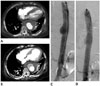
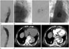
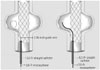
77-year-old woman who was admitted to our hospital due to coronary artery occlusive disease presented diffused abdominal and chest pain. A 6.5-cm saccular aneurysm with discontinuous intimal calcification of the descending thoracic aorta was found. She had increased C-reactive protein level at 19.02 mg/dL and leukocytosis (19.34 × 10³/uL), suggesting mycotic aneurysm (Fig. 1A). Broad-spectrum antibiotic therapy was started immediately. Due to her underlying disease and refusal to open surgery, TEVAR was performed with 30 mm diameter and 117 mm length stent-grafts (Valiant Captivia Medtronics, Minneapolis, MN, USA). After stent-graft placement, delayed filling of aneurysmal sac with reverse blood flow from the left seventh intercostal artery was found, suggesting type II endoleak (Fig. 1B, C). Follow-up postprocedural CT revealed the same findings (Fig. 1D). A secondary intervention was planned for this patient. One week after TEVAR, the patient had small amounts of hemoptysis. There was no definite increase in aneurysm size. Small amounts of hemoptysis suggested fistula between the aneurysm and the bronchus without underlying lung disease. Thus, thoracic aortography was performed. Thoracic aortography showed persistent type II endoleak related to retrograde filling of the aneurysmal sac with reverse blood flow from the left seventh intercostal artery (Fig. 2A). A small triangular space (free graft skirt) between a distal free graft wall of the stent-graft and the aorta was selected by a 5.0-Fr Davis-Berenstein catheter (Cook Incorporated, Bloomington, IL, USA) and a 0.35-inch hydrophilic J-tip wire (Terumo, Tokyo, Japan). The 5.0 F Davis-Berenstein catheter (Cook Incorporated, Bloomington, IL, USA) was then exchanged with a 5.0-Fr Straight catheter (Cook Incorporated, Bloomington, IL, USA) to keep a secure postion during a microcathter manipulation. A 2.0-Fr microcatheter (Progreat®; Terumo, Tokyo, Japan) with a 0.18-inch guide wire (Transend®; Boston Scientific, Boston, MA, USA) were advanced into the aneurysmal sac between the distal end of the stent-graft and the aortic wall by squeezing the very narrow space (Fig. 3). Cavitogram revealed opacification of the aneurysmal sac and efferent intercostal arteries (Fig. 2B, C). The aneurysmal sac was successfully embolized with a mixture of histoacryl (3 mL) and lipiodol (9 mL) at a dilution of 1:3 without selecting intercostal arteries (Fig. 2D). Postembolization angiography showed complete resolution of the endoleak (Fig. 2E). Follow-up CT angiography taken at 9 days after embolization showed that the size of the saccular aneurysm of the descending thoracic aorta was unchanged, but the endoleak was resolved (Fig. 2F, G). The patient did not complain of hemoptysis.
DISCUSSION
Endoleak is a significant predictive factor for outcome of thoracic aortic aneurysms treated by TEVAR (5). The incidence of endoleak after TEVAR ranges from 5% to 20%, which is similar to that after endovascular abdominal aortic aneurysm repair (EVAR) (1). Type II endoleaks are most commonly seen after EVAR. Type I and II endoleaks occur at similar rates after TEVAR (6). An accepted management method is aggressive endovascular repair of type I and III endoleaks along with observation for type II endoleaks (7). Collateral circulation in the chest involving the thoracic aorta is not so well developed compared to collateral vessels in the abdomen, making transarterial embolization of thoracic endoleaks quite difficult (6). To our knowledge, there is no consensus treatment option for type II endoleaks. There have been some reports on embolization of intercostal artery caused type II endoleaks by percutaneous sac puncture through lung parenchyma. However, a direct thoracic approach may involve transgression of the pleura and lung, which has a high risk of complications (6). In our case, embolization with a mixture of histoacryl (3 mL) and lipiodol (9 mL) was successfully performed with a transarterial approach in type II endoleak caused by reverse blood flow from the left intercostal artery. We placed a microcatheter in the aneurysmal sac between the distal end of the stent-graft and the aortic wall. This procedure may be difficult and unfeasible for some patients with anatomic limitations. Although there have been a few reports on transarterial embolization with liquid embolic agents for type II endoleaks after EVAR (8), there is no reported case like ours in the literature. The risk of spinal infarction and neulorogic deficits may contribute to this trend. Despite these drawbacks, we used liquid embolic materials to fill the aneurysmal sac and achieved successful embolization. Follow-up CT angiography confirmed the resolution of the type II endoleak caused by reverse blood flow from the left intercostal artery. This transarterial approach may be challenging but highly effective because the durability of endoleak embolization using the approach can be increased by selecting an actual endoleak with a microcatheter.
In conclusion, this is a report on successful transarterial embolization with hystoacryl in type II endoleak caused by reverse blood flow from the intercostal artery after TEVAR. Since there is no definite guideline for treating type II endoleaks after TEVAR, close and regular follow-up is needed for optimal treatment and good clinical outcome.
 XML Download