

 PDF
PDF ePub
ePub Citation
 Print
Print


INTRODUCTION
Leiomyosarcoma is the second most common primary retroperitoneal malignant tumor in adults, which commonly originates in the inferior vena cava (1). A leiomyosarcoma arising from the renal vein is particularly uncommon. After the first report of Varela and Garro in 1967, only about 30 cases have been reported in the published medical literature (2, 3). The renal vein leiomyosarcoma is grossly classified into three subtypes: completely extravascular; completely intravascular and mixed (1, 4). To the best of our knowledge, there have been only few reports detailing the imaging findings of renal leiomyosarcoma with both extra- and intravascular components (1, 4). We report an uncommon case of left renal vein leiomyosarcoma with combined components, focusing on the multidetector computed tomography (MDCT) and MR imaging findings with review of the literature. This case report was approved by the ethics committee at our institution and an informed consent was waived.
CASE REPORT
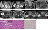
A 55-year-old female underwent an abdominopelvic MDCT to evaluate her epigastric pain for two weeks. The CT examination was performed using a 128-detector-row CT scanner (definition AS+, Siemens Medical Solutions, Forchheim, Germany). The enhanced CT scan of the abdomen showed a bi-lobed, heterogeneous, soft-tissue mass in the great vessel space and the left renal vein, measuring 9.5 cm in longest diameter (Fig. 1A, B). The extraluminal component abutted the uncinate process of the pancreas anteriorly, the aorta posteriorly and the portal vein superiorly (Fig. 1A, C). An expansile and branch-shaped intraluminal component occupied the lumen of the left renal vein, seen with a thin splaying of contrast medium (Fig. 1B). An abdominal MR imaging was performed for further evaluation on a 3.0T system (Magnetom Verio; Siemens Medical Solutions, Erlangen, Germany), using a body phased-array coil. The bi-lobed mass appeared isointense relative to the adjacent muscle on T1-weighted images (Fig. 1D). The extraluminal component showed a relatively low signal intensity and the intraluminal component showed an intermediate signal intensity with high-signal-intensity foci on T2-weighted images (Fig. 1E, F). Contrast-enhanced, fat-suppressed T1-weighted images showed a heterogeneous enhancement of the mass with a poorly enhancing portion, suggesting necrosis (Fig. 1G, H). The preoperative presumed diagnosis was either retroperitoneal sarcoma, including leiomyosarcoma or non-functioning neuroendocrine tumor with renal-vein thrombosis.
A complete excision of the tumor was performed. After the tumor and the affected segment of the left renal vein were removed, an 8 mm Dacron graft was used for end-to-end anastomosis between the left renal vein and the inferior vena cava. On gross examination, the extraluminal component measured 6.5-cm and the intraluminal component measured 3.5-cm in the longest diameter. The mass was gray and soft and showed hemorrhagic foci. The microscopic examination showed typical histologic features of leiomyosarcoma, including an interlacing fascicular pattern of proliferating spindle-shaped cells with nuclear atypia as well as a transition between the leiomyosarcoma and the intima of the renal-vein wall (Fig. 1I, J). These findings confirmed that the tumor arose from the wall of the renal vein. Immunohistochemical staining was positive for smooth muscle actin, appearing as brown (Fig. 1K).
DISCUSSION
Approximately 5% of leiomyosarcomas originate from the smooth muscles of the large blood vessels (5). The inferior vena cava is responsible for more than 50% of retroperitoneal leiomyosarcoma cases (2, 5). Primary leiomyosarcoma originating from the renal vein is quite uncommon with 50 to 60 years as prevalence peak age. It more commonly arises from the right renal vein compared to the left renal vein (2, 4). Long-term outcomes are closely related to tumor size and the feasibility of complete tumor resection (5).
On MDCT, a small leiomyosarcoma presents as a solid, soft-tissue density mass that is well-marginated and homogenous. However, a large mass can show necrosis and occasionally hemorrhage (6). Leiomyosarcoma shows low or intermediate signal intensity on T2-weighted MR images and low-intermediate signal intensity on T1-weighted MR images. It may present with several different degrees of enhancement and delayed enhancement compared to skeletal muscle (7). The signal intensity and enhancement seen on MRI depends on the amount of muscular and fibrous components and the extent of necrosis (8). In our case, both extraluminal and intraluminal components showed intermediate signal intensity on T2-weighted images with a portion of high-signal intensity observed inside the mass. The intraluminal component showed a fewer enhancement than the extraluminal component, probably because the intraluminal component contained more necrotic tissue and hemorrhagic foci. Unlike other retroperitoneal sarcomas that show a growth pattern which encases and narrows the renal vessel, neoplasms originating from the vessel wall show an intraluminal growth pattern or an intravenous propagation of the tumor (9). The presence of a solid and necrotic extravascular component with a contiguous intravascular enhancing portion is a more suggestive imaging finding of a retroperitoneal leiomyosarcoma (9).
A complete resection of the tumor and the involved renal vein using graft replacement is the treatment of choice. However, even following complete tumor resection, the five-year survival rate has been reported to be between 31% and 62% as more than half of these patients develop a tumor recurrence (4, 10).
In conclusion, the imaging findings of leiomyosarcoma are relatively non-specific. However, imaging findings such as an intraluminal component in the renal vein, heterogeneous enhancement due to combined necrosis and portions of low- or intermediate-signal intensity seen on T2-weighted MR images can suggest the diagnosis of a primary leiomyosarcoma of the renal vein.
 XML Download