

 PDF
PDF ePub
ePub Citation
 Print
Print


INTRODUCTION
The cytomegalovirus (CMV) is a DNA virus and a member of the herpes virus group. Symptomatic CMV infections usually occur in patients with severe immunodeficiency such as with acquired immunodeficiency syndrome, organ transplantation, hematologic malignancy, chemotherapy for malignancy, or steroid therapy (1). A CMV enterocolitis is the most common gastrointestinal manifestation in an immunocompromised patient. In immunocompetent hosts, symptomatic CMV enterocolitis is rare but may present with gastrointestinal bleeding, perforation or peritonitis. Radiologic findings of CMV enterocolitis are characterized by mural thickening of the small bowel and colon and frequently by segmental involvement and single halo enhancement pattern on CT (2). Recently, we treated a patient with CMV enteritis presenting with small bowel obstruction. The post contrast CT scan showed a focal narrowing at the jejunal loop and a thrombosis of the superior mesenteric artery (SMA). Herein, we describe a rare case of CMV manifestation with enterocolitis and the correlated imaging findings with pathologic findings.
CASE REPORT
A 51-year-old man with a 3-week-history of periumbilical pain visited our hospital. He lost 22 pounds body weight in one month. On physical examination, the patient appeared to be acutely ill with a blood pressure of 135/80 mm Hg, heart rate of 75 beats/min and a body temperature of 37℃. There was no specific previous medical history. The abdomen was distended with direct tenderness in the periumbilical area. The white blood cell count was 12.01 × 10³/µL and the C-reactive protein was 4.54 mg/dL, showing an inflammatory condition.
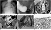
A plain radiograph of the abdomen showed dilated small bowel loops (Fig. 1A). A double contrast small bowel follow-through showed a beaklike narrowing with passage disturbance of contrast material without mucosal irregularity at the distal jejunum (Fig. 1B). Also the proximal portion of jejunal loops was dilated.
The contrast-enhanced computed tomography (CT) scans showed a mild focal luminal narrowing without mass at the distal jejunum and a relatively long segmental partial thrombosis in SMA (Fig. 1C). Due to relatively long segmental SMA thrombosis (Fig. 1D), we diagnosed a benign stricture associated with infection such as enteritis or focal ischemic change as there was no visible mass, mural thickening, edema or perienteric infiltration and a mucosal enhancement was unclear. But we could not exclude the possibility of small bowel malignancy such as adenocarcinoma because of the patient's age and significant weight loss. Also we could not recommend a capsule endoscopy due to the persistent symptom of bowel obstruction. A segmental resection of the small bowel was done and a segmental stricture was pathologically confirmed (Fig. 1E). The immunohistochemical stain was consistent with a CMV infection (Fig. 1F).
DISCUSSION
CMV is a herpes virus that usually does not cause symptoms in an immunocompetent host. However, a CMV infection can present as asymptomatic viremia or CMV syndrome with viremia and symptoms including fever and malaise, or even as tissue-invasive disease, such as colitis, hepatitis, pneumonitis, myocarditis, meningoencephalitis and rarely retinitis in immunocompromised subjects. The involvement of the gastrointestinal tract is a relatively common manifestation of a tissue-invasive CMV disease (1). CMV can cause lesions throughout the gastrointestinal tract from the mouth to the anus. The lesions are various, but ulcerative lesions causing mainly colitis are most common. Other pathological lesions caused by CMV infection of the gastrointestinal tract could be perforations, hemorrhagic proctocolitis, inflammatory pseudotumor, appendicitis, toxic megacolon or pneumatosis intestinalis. Less than 10% of the CMV gastroenteritis cases show small bowel gastroenteritis (2, 3). In an immunocompetent patient, CMV enteritis can cause symptoms like in an immunocompromised host. A focal small bowel stenosis like in the presented case is a rare finding of CMV enteritis.
In one study were the CT findings of CMV enterocolitis in immunocompetent hosts similar to those reported in patients with acquired immunodeficiency syndrome (4). The most common CT finding of CMV enterocolitis was segmental and concentric mural thickening involving the colon was present in most patients and the small bowel was involved in approximately 30%. Isolated enteritis was noted in one patient of 12 patients and a focal involvement of rectum was seen in one case also. A stenosing and ulcerative CMV infection of the colon resembled a neoplasm clinically, macroscopically and radiologically. A case of gastric antral obstruction due to mass has also been described caused by CMV infection (5, 6).
We considered a malignant neoplasm as one of the possible differential diagnosis because of the focal involvement of the small bowel. However, there was no definite radiologic evidence of neoplasm such as mass-like lesion, enlargement of mesenteric lymph node or desmoplastic reaction. Adhesion could be a possible diagnosis based on the fact that the lesion showed a dramatic luminal narrowing with no visible cause. So, we put more weight on small bowel obstruction caused by inflammatory condition or post ischemic change.
Although the mechanism remains unclear, a causative association between CMV infection and thromboembolic disease has been proposed, most frequently described in the context of immunosuppression after organ transplantation (7). The proposed mechanism in most instances is that of a thrombotic microangiopathy. It is suggested that viruses are able to alter the phenotype of endothelium from anticoagulant to procoagulant, thus promoting the adhesion of neutrophils and platelets (8). But CMV induced vasculopathy and thrombosis are rare conditions. The few published reports on these conditions focus either on immunocompromised transplant recipients who are receiving high-dose immunosuppressive agents or on HIV-infected patients (9). There are few reports involving immunocompetent adults in whom no hemostatic abnormalities can be found similar to our patient (10). Our case showed a CMV enteritis, presenting as a small bowel obstruction accompanied by SMA thrombosis in an immunocompetent individual with no underlying disease. Regarding the superior mesenteric artery thrombosis, recent studies showed that the cytomegalovirus can accelerate a vascular disease by inflammation and smooth muscle cell migration from the vessel media to the intima and by proliferation that culminates in vessel narrowing. In our case, distal SMA different from proximal SMA thrombosis was opacified on the contrast enhanced CT and we hypothesized the SMA thrombosis occurred at a significant time point before so that peripheral branches could be retrogradely perfused by collaterals. Although our treatment options could include antibiotics or heparinization, we performed a segmental resection of the small bowel to rule out the possibility of malignancy. The histology confirmed the CMV infection. The significance of SMA thrombosis was not fully evaluated in this study, but we supposed the SMA vasculopathy as a result of the CMV infection.
In conclusion, we experienced an extremely rare case with CMV small bowel involvement and SMA at the same time. It was successfully treated with surgical intervention and complimentary
antiviral medication.
 XML Download