

 PDF
PDF ePub
ePub Citation
 Print
Print


INTRODUCTION
Primary hepatocellular carcinoma (HCC) is one of the most common cancers worldwide with a high mortality rate. This malignancy occurs more often in men than women, with the highest incidence rates reported in East Asia (1). Spontaneous regression of malignant tumors is a rare phenomenon, and estimated to occur once in 60000-100000 cancer patients (2). Fewer than 70 cases are reported in the literature, and the reasons behind these occurrences remain unknown (3). To our knowledge, there is no case of spontaneous regression after liver biopsy. Furthermore, cases of spontaneous regression of large HCC are very rare. There are only two case reports on the recurrence of HCCs after spontaneous regression. We report on one patient with a 7.5 cm HCC tumor that regressed spontaneously after ultrasound (US) guided liver biopsy and re-grew after stable disease during a 28-month period.
CASE REPORT
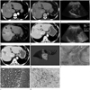
A 64-year-old female visited our hospital with intermittent abdominal discomfort. The patient had a history of chronic hepatitis B infection without treatment for 20 years, as well as congestive heart failure. Her vital signs were stable and there were no remarkable physical findings. Hemogram and liver enzyme were normal. Enzyme immunoassay was positive hepatitis B surface antigen and extracellular hepatitis B antigen, and negative for hepatitis B surface antigen and hepatitis C virus. Hepatitis B virus DNA titer was 14546 IU/mL (49601 copies/mL). The level of alfa-fetoprotein (AFP) and protein induced by vitamin K antagonist (PIVKA II) was elevated to 95.62 ng/mL (normal range, 0-15 ng/mL) and 903 mAU/mL (normal range, 0-40 mAU/mL) at admission, respectively. Contrast-enhanced computed tomography (CT) of the abdomen showed an ill-defined heterogeneous enhancing mass in the arterial phase with a maximal diameter of 7.5 cm in Couinaud's segmental V/VIII of the liver (Fig. 1A). This mass showed washing-out of the contrast medium in the portal phase and delayed phase (Fig. 1B). The initial US showed a similar-sized heterogeneous hypoechoic mass with a rim halo (Fig. 1C). After 2 weeks, an US guided liver biopsy revealed relatively discrete inflammatory lesions composed of chronic inflammatory cell infiltration, loose or hyalinized fibrosis with vascular proliferation, and focal coagulation necrosis. Although the biopsy did not reveal malignancy, radiologic and laboratory findings of the tumor were evident as HCC. So, the radiologists and clinicians agreed to follow-up closely without anti-cancer therapy. After 1 week, follow-up magnetic resonance imaging (MRI) of the abdomen revealed that the tumor was decreased in size to 3.3 × 3.5 cm (Fig. 1D). After 4 months, the serum PIVKA II level had decreased to 31 mAU/mL and the follow-up CT (Fig. 1E) and band US (Fig. 1F) showed a further decrease in size to 2.4 × 1.8 cm. The tumor showed partial peripheral nodular enhancement in the arterial phase and contrast washout in the delayed phase on CT. Almost every 6 months, repeated abdominal CT scan showed no gross interval change of this lesion during 22 months. Serum AFP level was normal, but serum PIVKA II level had gradually elevated. In the process, a follow-up CT of the abdomen performed 28 months after the initial CT showed a size increase of up to 3.7 × 2.3 cm in S8, and elevation of serum PIVKA II to 1143 mAU/mL (Fig. 1G). S8 segmentectomy of the liver showed a confluent multinodular mass associated with massive necrosis, focal hemorrhage, and mixed loose or myxoid fibrosis and thick walled biliary trees (Fig. 1H). Microscopically, along with the margins of necrotic nodules, variable portion of poorly differentiated HCC cells were present, which were mostly mixed with inflammatory cell infiltration (Fig. 1I, J). Some of the poorly differentiated HCC cells revealed a CK19 expression (Fig. 1K).
DISCUSSION
Spontaneous regression of a malignant tumor was defined by Everson and Cole as a partial or complete involution of a malignant tumor, without the application of any specific therapy. The incidence of spontaneous regression was estimated to be one per 60000-100000 cases of malignancy (4).
The mechanism of spontaneous regression of HCC is not clear. Suggested factors include alcohol withdrawal, androgen withdrawal, intake of herbal medicine, stimulation of cytokine production, and fever (5,6,7,8,9). Among the various factors, ischemia is the most common etiology of spontaneous regression (10). Rapid growth, arterioportal shunt, formation of a thick capsule, and portal vein thrombosis restrict the blood supply to the tumor, which can result in tumor necrosis and regression of HCC (10). In our case, spontaneous regression of HCC occurred after liver biopsy. Therefore, we speculate that the liver biopsy injured the HCC feeding vessel and caused ischemic damage, which lead to the partial spontaneous regression of large HCC.
Two points should be taken into consideration for the size reduction of tumor in our case. First, there could have been measurement errors because different imaging modalities were used to evaluate HCC before and after liver biopsy; CT at initial diagnosis and MRI 1 week after biopsy. However, the results of follow-up CT, US, and tumor marker 4 months after biopsy demonstrated a meaningful size reduction of tumor. Second, in terms of the absence of tumor cells in the biopsied tissue, the initial tumor size can be overrated due to peritumoral inflammation. Although the initial US guided liver biopsy showed relatively discrete inflammatory lesions featuring chronic inflammatory cell infiltration without cancer cell infiltration, segmentectomy showed that variable populations of poorly differentiated HCC mostly mixed with inflammatory cell infiltration was present, along with the margins of necrotic nodule. We assume that the US guided liver biopsy was incorrectly targeted at the inflammatory cell infiltration site. Histopathologic findings of viable portion of HCC along the margin of necrotic nodule were correlated with that of radiologic findings of the tumor that showed peripheral enhancement in the arterial phase and washout in delayed phase at the repeated abdominal CT scans.
Cases of spontaneous regression of HCC are rare. Furthermore, spontaneous regression after liver biopsy has hitherto not been reported. We hope that this case will help further the understanding of the etiology of spontaneous regression and to develop treatment strategies for HCC.
 XML Download