

 PDF
PDF ePub
ePub Citation
 Print
Print


INTRODUCTION
The aberrant internal carotid artery (ICA) in the middle ear is a rare, but important vascular anomaly of the temporal bone. The clinical diagnosis of an aberrant ICA is often difficult, because signs and symptoms such as conductive hearing loss, pulsatile tinnitus and vertigo are nonspecific. The aberrant ICA is frequently confused with otosclerosis, glomus tumor and other vascular malformations such as dehiscent jugular bulb, hemangioma and aneurysm (1, 2). A misdiagnosis of this anomaly could have serious consequences. An excessive aural bleeding during a myringotomy or tympanotomy is a life-threatening complication (3). Although the identification became easy with the temporal bone computed tomography (CT), it is not unusual for this anomaly to be discovered during middle ear surgery. Our literature review yielded 78 cases of aberrant ICA in the middle ear, with 11 of them presenting with a persistent stapedial artery. Herein, we report a case of aberrant ICA in the middle ear and describe the clinical and radiological features of this vascular anomaly.
CASE REPORT
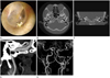
A 38-year-old woman visited our hospital due to tinnitus and hearing difficulties of the left ear that had started 5 years ago. During otoscopy, an anteroinferior bluish pulsating mass was seen in the tympanic space (Fig. 1A). The audiometry showed a hearing loss of conductive type in the left ear. High resolution temporal bone CT scanning and CT carotid angiography were performed and showed a left-side aberrant ICA with a bony dehiscence of the carotid canal (Fig. 1B). The left ICA was seen entering the tympanic cavity through the markedly enlarged inferior tympanic canaliculus (Jacobson canal) (Fig. 1C), crossing the cochlear promontory and projected into the tympanic space (Fig. 1D). The native vertical portion of the petrous carotid canal and the foramen spinosum were absent. The magnetic resonance angiography (MRA) showed a reduced diameter of the tympanic ICA. The vertical segment of the carotid artery was laterally located to a line drawn vertically through the vestibule. An aplasia was revealed of the A1 segment of the left anterior cerebral artery (ACA) (Fig. 1E).
DISCUSSION
Normally, the ICA enters the petrous bone medial to the styloid process via the carotid canal. The initial vertical segment is separated from the tympanic cavity by a thin plate of bone. The ICA then turns anteriorly to lie inferior and posteromedial to the eustachian tube, traverses the foramen lacerum and enters the medial cranial fossa.
Rarely, the ICA takes an aberrant course. Several hypotheses have been considered concerning the genesis of aberrant ICA. Lasjaunias and Santoyo-Vazquez (4) hypothesized the alternate blood flow theory that the C1 portion of the ICA involutes owing to the persistence of the pharyngeal artery system and as a consequence, an anomalous course develops with blood flowing via the ascending pharyngeal artery to the enlarged inferior tympanic artery with retrograde flow through the caroticotympanic vessels into the horizontal segment of the ICA. This theory may explain the radiological features with an enlargement of the inferior tympanic canaliculus, the presence of a mass like lesion found in the anterior hypotympanum and the absence of the vertical portion of the ICA.
The clinical diagnosis of an aberrant ICA appears as difficult because signs and symptoms such as pulsatile tinnitus, conductive hearing loss and a pulsatile tympanic mass in the anteroinferior area are often nonspecific or absent (5, 6). The results of a conductive hearing loss component from an audiometric evaluation may be attributed to a malleus or incus blockage. Those findings could be regarded as otosclerosis, glomus tumor or other vascular malformation. However, a tympanic mass due to an aberrant ICA looks different from a glomus tumor and dehiscent jugular bulb: anterior, pulsatile and white or rosy (1, 2). Thus, a mostly asymptomatic aberrant ICA will be diagnosed during middle ear surgery (7). A temporal bone CT should be performed before any middle ear surgery in order to avoid a surgical injury due to misdiagnosis. On CT scan, an aberrant ICA is identified by an ICA that runs adjacent to the jugular bulb, in a posterior position and with a reduced diameter, an enhancing mass in the hypotympanum, a deficient bony plate along the tympanic portion of the ICA, an enlargement of the inferior tympanic canaliculus and the absence of the vertical segment of the carotid canal (2). The MRA can be used as an additional tool if it a definitive diagnose is not possible with a CT scan only (6). It provides an excellent visualization of the intracranial and extracranial circulation. In this case, the MRA showed a reduced diameter of the tympanic ICA, the absence of the vertical segment of the carotid canal and a aplasia of the A1 segment of the ACA on the same side. All of these anomalies were located on the same side and probably may have a common cause. This common cause is most likely a maldevelopment of the vascular network (1).
The knowledge about this rare entity is essential for a clinician, because an accidental injury after myringotomy or in case of misdiagnosis with another vascular tumor may lead to disastrous consequences. Most authors recommend a conservative approach in case of an asymptomatic and proven aberrant ICA (7, 8), but Ruggles and Reed (9) advocated a surgery to relieve the patient of troublesome symptoms and to prevent a possible destruction of the middle ear structures and formation of an aneurysm.
The knowledge about the aberrant ICA in the middle ear is essential for clinicians, because a misdiagnosis of this anomaly could lead to serious consequences such as excessive aural bleeding or vascular occlusion. All masses in the middle ear, especially pulsating masses, should be studied by imaging methods such as CT and MRA.
 XML Download