

 PDF
PDF ePub
ePub Citation
 Print
Print


INTRODUCTION
Placement of a gastroduodenal stent is an important and effective palliative treatment in patients with unresectable gastroduodenal obstruction (1, 2). The incidence of complications from gastroduodenal stenting ranges from 17% to 25% (3, 4). Reported complications include stent obstruction caused by tumor ingrowth or overgrowth, or by food impaction, stent migration, biliary obstruction, perforation, and bleeding (5, 6). Among these complications, acute biliary obstruction is uncommon, but has a high mortality rate (1, 5). We report two cases of acute obstructive jaundice after placement of an uncovered stent in the pylorus and duodenum was managed with biliary stent placement.
CASE REPORT
Case 1
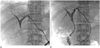
A 50-year-old male was admitted to the hospital complaining of right upper quadrant pain and vomiting. Initial abdominal computed tomography (CT) showed a stomach mass in the pylorus, narrowing the lumen, and multiple enlarged perigastric lymph nodes (LNs). Based on the CT image, advanced gastric cancer was diagnosed as stage IIIB. The patient was not a candidate for surgery because of the advanced stage of his stomach cancer. Therefore, a self-expandable, uncovered type pyloric stent (18 mm × 10 cm; Bona stent, Sci-Tech Inc., Seoul, Korea) was placed successfully for palliative treatment. After the procedure, the patient was able to tolerate a low residue diet without vomiting and showed a normal total bilirubin level. Five days after placement of the stent, the patient was referred for management of an elevated total bilirubin level (6.6 mg/dL) and jaundice. Ultrasonography showed diffuse dilatation of intrahepatic ducts from the proximal common bile duct (not shown here). Percutaneous biliary drainage (PTBD) was performed via the left lateral segmental duct under ultrasonographic guidance. A tubogram revealed blockage of the common bile duct, which was interpreted to indicate acute extrahepatic duct compression by the expanded pyloric stent and multiple enlarged perigastric LNs (Fig. 1A). Six days after PTBD, we placed a biliary stent (10 mm × 7 cm; Hercules, S&G Biotech, Seongnam, Korea) in the common bile duct. Follow-up tubogram after the biliary stent insertion revealed good passage of contrast media via the biliary stent into the duodenum (Fig. 1B), and the total bilirubin level decreased to 1.38 mg/dL and remained normal for 118 days.
Case 2
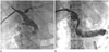
A 76-year-old female was referred to our hospital with complaints of abdominal discomfort and recurrent vomiting. Gastroduodenal endoscopy revealed advanced carcinoma in the second and third portion of the duodenum. We placed an uncovered duodenal stent (20 mm × 14 cm; Hanaro stent, SolcoIntermed, Seoul, Korea) for palliation. After duodenal stent placement, the patient tolerated a soft diet and the total bilirubin level was within normal ranges. Six days after duodenal stent placement, the patient was referred again because the total bilirubin levels had rapidly increased (5.1 mg/dL). CT showed diffuse dilatation of extra- and intra-hepatic ducts (not shown here). PTBD was performed via the right posterior segmental duct and a tubogram showed complete obstruction at the Ampulla of Vater with dilatation of the biliary tree (Fig. 2A). Acute biliary obstruction was assumed to result from compression of Ampulla of Vater by expansion of the duodenal stent and displaced duodenal cancer. To manage the obstruction, a biliary stent (10 mm × 7 cm; Hercules stent, S&G Biotech, Seongnam, Korea) was placed through the mesh of the duodenal stent after pre-stent balloon dilatation with a 6 mm × 4 cm balloon. A follow-up tubogram, 3 days after placement, of the biliary stent showed good passage of contrast media into the duodenum and jejunum through the biliary and duodenal stents (Fig. 2B). Five days later, the total bilirubin level was also normalized (0.8 mg/dL). The patient was monitored for 135 days and remained asymptomatic through that time.
DISCUSSION
Malignant gastroduodenal obstruction often coincides with biliary obstruction, both of which cause severe symptoms: vomiting, nausea, anorexia, and weight loss in cases of gastroduodenal obstruction and jaundice, pruritus, and cholangitis in biliary obstruction (7). Because most of these cases are not candidates for curative surgery, metallic stent insertion provides a safe, effective and feasible alternative palliation (1, 3). However, acute biliary obstruction secondary to placement of a gastroduodenal stent has been reported to occur infrequently, though its rate of incidence is not defined (1, 5, 8). The suggested mechanism is that the gastroduodenal stent covers the papilla. Therefore, biliary obstruction after placement of a gastroduodenal stent has been found to occur when a covered stent is placed over the papilla (1, 2, 9). Previous studies (2, 8) recommended that an uncovered stent should be used or biliary decompression should be mandatory prior to placement of the covered stent in the second portion of the duodenum. However, the authors think that further studies are needed to evaluate the exact causes of acute biliary obstruction after gastroduodenal stent placement and to extablish a standard treatment protocol.
Profili et al. (10) reported delayed biliary obstruction in a benign stricture patient 3 months after placement of an uncovered duodenal stent caused by stent-induced chronic inflammation of the duodenal wall. However, the acute secondary biliary obstructions in our cases occurred, within 6 days of uncovered stent placement. Therefore, we feel biliary obstruction resulted from compression of the bile duct by an expanded gastroduodenal stent and displaced malignant tumor or enlarged LNs. Furthermore, in our first case, the biliary obstruction occurred at the level of the mid-common bile duct. This type of secondary biliary obstruction appears to be unusual. In our cases, percutaneous biliary stent placement was successful, and total bilirubin was normal during the follow-up period, even though the biliary stent had a smaller diameter and lower radial force than the gastroduodenal stent.
In conclusion, we report two cases of acute biliary obstruction after placement of uncovered stents in the pylorus and duodenum. In these cases, percutaneous biliary drainage followed by biliary stent placement resolved the obstruction.
 XML Download