

 PDF
PDF ePub
ePub Citation
 Print
Print


INTRODUCTION
The most common giloma in patients with neurofibromatosis type 1 (NF1) is the pilocytic astrocystoma (1, 2). It commonly involves the optic tract and hypothalamus, in the order of frequency (2). However, the pilocytic astrocytoma is rarely found in other intracranial regions of NF1 patient (3).
Also, pilocytic astrocytoma usually has a classic imaging manifestation of a solitary, cyst-like mass with an strong contrast-enhancing mural nodule (4). Therefore, while the isolated pilocytic astrocytoma in NF1 is being well-described and frequently reported, multiple pilocytic astrocytomas in an individual patient is a less common form of manifestation. There has only been one single publication on the multiple involvements of pilocytic astrocytoma, which was located within the cerebellum of NF1 patient recorded in the United States in 2007 (2). According to our knowledge, no such case has been described since, thus, herein, we report a case of pilocytic astrocytoma presenting with only solid, multiple pilocytic astrocytoma in the cerebellum of the NF1 patient.
CASE REPORT
A 10-year-old male patient with a known history of NF1 was originally diagnosed from the presence of axillary freckling, multiple café-au-lait macules (> 6), and an affected first-degree relative presented with 1 month of headache. He underwent computed tomography (CT) and magnetic resonance imaging (MRI) of the brain in another hospital, and was found with multiple cerebellar masses. He visited our hospital for further evaluations and treatments.
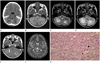
His initial symptoms only included headaches, and there were no other neurological symptoms. We reviewed the external CT, which showed bilateral low densities with central iso- or high-density in both cerebellums (Fig. 1A). MRI found multiple well-demarcated nodular contrasts enhancing solid lesions in both cerebellar hemispheres with heterogenous low-signal intensity on T1-weighted images, and heterogenous high-signal intensity on T2-weighted images (Fig. 1B-E). Perilesional vasogenic edema surrounding these multiple enhancing solid lesions were also found, which did not cause obstructive hydrocephalus. Generally, findings known as hamartomas or heterotopias (1), typically found in NF1 patients and presented as iso-signal intensities on T1-weighted images, and high-signal intensities on T2-weighted images without contrast enhancements, were found in the frontal subcortex, the right periventricular white matter, the posterior limb of right internal capsule, both the basal ganglia, the right thalamus, pons and the left superior cerebellum (Fig. 1F).
For the histological analysis, a left suboccipital craniotomy and an open biopsy was performed for one of the enhancing lesions in left cerebellar hemisphere. Pathology was consistent with low graded pilocytic astrocytoma (World Health Organization grade I), according to standard features of biphasic appearance together with loose glial components of multifocal myxoid changes and more compact piloid tissues with elongated nuclei (Fig. 1G). Cytologically, the tissues were positive for glial fibrilary acidic protein.
The patient was discharged without neurological deficits, and after 2 months, he underwent a gamma-knife surgery for multiple enhancing cerebellar solid masses. He was discharged after displaying no remarkable postoperative complications.
DISCUSSION
NF1, formerly known as von Recklinghausen's disease or peripheral neurofibromatosis, is a relatively common (incidence of 1 in 3000-4000 births) autosomal dominant disorder, and is related to mutations in chromosomes 17 (17q11.2) which are encoded for the protein neurofibromin (1).
Although occurring less frequent than peripheral tumors, the central nervous system tumors are important for NF1 patients because they may lead to major morbidity and mortality rates (1, 2). The most common intracranial tumors are optic gliomas, which occur in about 5-15% of NF1 patients. Brain gliomas are observed in about 1-3% of the patients, and the most common glioma in NF1 is pilocystic astrocytoma (1, 2).
The association of pilocytic astrocytoma with NF1 has been well-documented (3, 4). For example, the cytogenetic analysis of pilocytic astrocytomas have detected losses of genetic materials involving the long arm of chromosomes 17 (17q) near the same locus for the NF1 tumor suppressor genes. Furthermore, the lack of expression on an NF1 gene product, the neurofibromin, has been documented in NF1-associated pilocytic astrocytomas (3, 4). These findings have fueled the speculations that NF1 tumor suppressor genes are linked with the expressions of pilocytic astrocytoma.
Pilocytic astrocytoma in NF1 patients typically involves the optic nerves or chiasms, which comprise up to about 14% to 15% of all tumors associated with NF1 (1-5), and 3.7% of brain stem tumors (1, 3, 5), but the actual percentages vary among different studies. Pilocytic astrocytoma is rarely found in other intracranial regions of NF1 patients. Cerebellum is an uncommon location for tumors in patients with NF1, and rarely surpasses a prevalence of 1% (3).
On the other hand, regardless of associations with NF1, the pilocytic astrocytoma is generally the most common primary brain tumor in children, comprising around 85% of all cerebellar astrocytomas and 10% of all cerebral astrocytomas within this age group (4). Most lesions arise from the cerebellum, the optic nerve and chiasm, or the region of the hypothalamus-thalamus, and occur within or near the midline (4) sections.
The clinical presentations of pilocytic astrocytoma vary with its site of origin. Symptoms, commonly found in cerebellar pilocytic astrocytoma, include headaches, vomiting, gait disturbances, blurred visions, diplopia, and neck pains (4, 6). Clinical signs usually include hydrocephalus, papilledema, truncal ataxia, appendicular dysmetria, head tilt, sixth nerve palsy, and nystagmus (4, 6).
The treatment of pilocytic astrocytoma also varies according its origin. Surgical resection is considered the treatment of choice for cerebellar pilocytic astrocytomas and is generally regarded as curative after gross removals of the tumor (4, 6). For lesion locations involving difficult approaches, the stereotactic resection may be used (4). Overall, the prognosis for patients with a pilocytic astrocytoma is excellent, usually with a 94% of 10-year survival rates and a 79% of 20-year survival rates (4).
On CT imaging, most pilocytic astrocytomas have well-defined borders with either a round or oval shape, smaller than 4 cm in size, cyst-like features, smooth margins, and occasional calcifications (4). In a study by Coakley et al. (7), most tumors (82% in one series) were located near the ventricle, and almost all of them (94%) showed intense enhancements on post-contrast images which were obtained after intravenous administration of contrast materials (4).
Numerous studies have described four predominant imaging patterns of pilocytic astrocytoma on MRI-I: mass with a non-enhancing cyst and an intensely enhancing mural nodule, II: mass with an enhancing cyst wall and an intensely enhancing mural nodule, III: necrotic mass with a central nonenhancing zone, and IV: predominantly solid mass with minimal to no cyst-like component (4, 6). Most studies have also shown that approximately two-thirds of all pilocytic astrocytoma demonstrated the typical imaging manifestation of a cyst-like mass with strong contrast-enhancing mural nodule. The degree of surrounding vasogenic edema are being diminished, which the expectation for a tumor with low biological activity (4).
However, there are occasional atypical imaging manifestations of pilocytic astrocytoma. More rarely, the multiple involvement of pilocytic astrocytomas may be presented within an individual patient (2, 8, 9). Such phenomenon has only been reported few times before-pilocytic astrocytoma in the multiple compartment (supra/infratentorial and/or spinal cord) in a patient without NF1 (8) and in same cerebral hemisphere associated with the NF1 patient (9). Also, multiple pilocytic astrocytomas in one cerebellar hemisphere of a patient with NF1 have been reported once by Dunn et al. (2). These case reports are meaningful due to the lack of literature for multiple types of pilocytic astrocytomas or posterior fossa tumors in patients with NF1.
Our case is similar to the one reported by Dunn et al., which described multiple solid and cystic type pilocytic astrocytomas in the cerebellum of NF1 patient. However, our case is different in that there are only enhancing solid types of multiple pilocytic astrocytomas in the cerebellum, which is a less common manifestation rather than a typical manifestation of cyst-like mass with an strong contrast-enhancing mural nodule type, such as the report by Dunn et al. Thus, we believe our case to be more of a rare form of multiple pilocytic astrocytomas in the cerebellum.
In conclusion, we report a surgically and pathologically proven case of pilocytic astrocytomas which is comprised of multiple, solid enhancing nodules in both cerebellar hemispheres of the NF1 patient. Radiologists should expand the spectrum of presentation for patients with NF1 and consider multiple pilocytic asctorcytoma as a differential diagnosis when encountering multiple enhancing lesions that involve both cerebellar hemispheres in the NF1 patients.
 XML Download