

 PDF
PDF ePub
ePub Citation
 Print
Print


INTRODUCTION
Brodie's abscess, a localized type of subacute or chronic osteomyelitis, may be difficult to differentiate from benign or malignant bone tumors (1). The "penumbra sign" on an unenhanced T1-weighted image is a well-known characteristic of Brodie's abscess. It is seen on T1-weighted MR images as a rim lining the abscess cavity with a higher signal intensity than that of the main abscess. It is considered to be extremely helpful for discriminating subacute osteomyelitis from other bone lesions, although it is not pathognomonic.
We describe a case of histopathologically confirmed primary diffuse large B-cell lymphoma of the distal tibia with a positive penumbra sign on MRI. To our knowledge this is the first report of a primary bone lymphoma that shows the finding of a penumbra sign on an MRI.
CASE REPORT
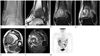
A 50-year-old woman presented with a 3-month history of left ankle pain. As the pain worsened, she visited a local clinic and was treated with non-steroid anti-inflammatory drugs. But the pain did not improve. She was referred to our hospital for further investigation. Physical examination revealed tenderness and redness over the medial aspect of the left ankle with preservation of the full range of motion. She had diabetes mellitus and had received oral hypoglycemic agents for 8 years. There was no fever or other systemic complaints. Laboratory evaluation showed a normal white blood count of 6.5 × 10³/mm3 and a normal C-reactive protein level of less than 0.3 mg/dL. Radiographs of the left ankle (Fig. 1A) showed an ill-defined intramedullary osteolytic lesion involving the distal metaphysis and epiphysis of the tibia. The overlying cortex was intact, and there was no visible periosteal reaction or matrix calcification. Further evaluation using MRI demonstrated a 2.9 × 2.4 × 3.2 cm sized lesion that had a center that was isointense to skeletal muscle on T1-weighted images (Fig. 1B, E) and that showed hyperintensity on T2-weighted spectral attenuated inversion recovery (SPAIR) images (Fig. 1C). There was a thin sclerotic rim around the center and a dot of dark signal intensity on the T1 and T2-weighted images resembling a bony sequestrum. A small enhancing focus was seen in the center, but most of the center showed no contrast enhancement (Fig. 1D, F). The center was surrounded by two layers of different MR signal features and an outermost halo of bone marrow edema. The inner layer demonstrated slightly higher signal intensity than the center on T1-weighted images (Fig. 1B, E) and high signal intensity on T2-weighted SPAIR images (Fig. 1C). It showed strong gadolinium enhancement on contrast enhanced T1-weighted images (Fig. 1D, F). The outer layer showed hypointensity on T1-weighted images (Fig. 1B), mixed high and low signal intensity on T2-weighted images (Fig. 1C), and mild enhancement following contrast medium administration (Fig. 1D, F), consistent with reactive sclerosis and edema. There was also evidence of cortical permeation and mild active periostitis on the MRI (Fig. 1F).
A preliminary diagnosis of subacute osteomyelitis was made because the MRI findings were most consistent with Brodie's abscess. We considered giant cell in the differential diagnosis of the tumor because the lesion involved the epiphysis and metaphysis simultaneously. The possibility of a malignancy also could not be ruled out.
The patient underwent curettage of the lesion. The curetted materials were yellow-white tissue fragments without pus. Histopathological examination of the curettage specimen showed extensive coagulative necrosis surrounded by a diffuse sheet of large lymphoid cells with round to slightly indented vesicular nuclei containing fine chromatin. There were two to four nuclear membrane-bound nucleoli, frequent mitotic figures and tingible body macrophages. The cytoplasm was not abundant and it was pale eosinophilic to amphophilic in appearance (Fig. 2A). Immunophenotypical analysis detected the expression of B-correlated antigens: CD20 (Fig. 2B) and CD79A, Bcl-2, Bcl-6, and MUM1. EBER mRNA in situ hybridization was negative. The histopathological diagnosis was diffuse large B-cell lymphoma, a centroblastic variant, involving the bone marrow space. Nine days after the surgery, the patient underwent a positron emission tomography/CT (PET/CT) scan. There was no evidence of other primary sites or lymph node involvement (Fig. 1G).
The patient was referred to oncology, and underwent chemotherapy. After completion of six courses of the R-CHOP regimen (rituximab, cyclophosphamide, doxorubicin, vincristine and prednisone), follow-up studies of the MRI and PET/CT demonstrated no evidence of recurrence.
DISCUSSION
Bone tumors can be misdiagnosed as other non-neoplastic conditions including infections on clinical and radiologic examination (2). And there may be considerable difficulty in distinguishing subacute osteomyelitis from other benign and malignant bone lesions, resulting in delayed diagnosis and treatment. For example, there are some case reports of bone tumors such as osteoid osteoma and chondrosarcoma mimicking Brodie's abscess (2, 3). Similarly, malignant lymphomas of the bone mimicking osteomyelitis or abscess have been documented as case reports (4, 5). However, to our knowledge, there is no previously reported case with the characteristic MR appearance of the "penumbra sign" mimicking Brodie's abscess.
The "penumbra sign" means the inner ring of the "target" that is characteristically seen on T1-weighted MR images as a discrete peripheral zone of marginally higher signal intensity than the abscess cavity. It is considered to be a characteristic MRI feature of Brodie's abscess with a high degree of specificity (1). Grey et al. (6) reported the sensitivity, specificity, accuracy, positive predictive value and negative predictive value of the penumbra sign in diagnosing subacute osteomyelitis to be 75%, 99%, 99%, 92% and 99%, respectively. However, this sign is not pathognomonic; it has also been reported in eosinophilic granuloma, benign cystic bone lesions and bone tumors such as osteoid osteoma (2) and chondrosarcoma (3).
The "target" appearance of Brodie's abscess on MRI is comprised of four separate layers: a center, an inner ring, an outer ring and a peripheral halo (7). The center represents the abscess cavity with a high protein content. The inner ring is composed of granulation tissue that appears isointense or mildly hyperintense to muscle on T1-weighted and shows intense enhancement after contrast administration due to its rich vascularity. The outer ring is composed of a zone of reactive sclerosis and the peripheral halo represents bone marrow edema.
However, in the curettage specimen from our case, there was no evidence of abscess formation. There were several hemorrhagic fragments surrounded by fibroblasts and they may have composed the central non-enhancing portion of the lesion. The rest of the curetted fragments were mainly composed of lymphoma cells. Primary bone lymphoma is one of the rare primary bone malignancies, accounting for 5% of all primary bone tumors (8, 9), and the majority of primary bone lymphomas is non-Hodgkin's lymphoma, especially diffuse large B-cell type (9). Common anatomical sites for primary bone lymphomas are the long bones such as the femur (25%), the tibia and the humerus, in decreasing order of incidence (8, 10). Less commonly, they involve the flat bones or the axial skeleton such as the pelvic bone and the vertebral column. Common symptoms are pain, local swelling and a palpable mass (9).
Patterns of radiographic findings in primary bone lymphomas have varied from a near-normal lesion to a focal geographic lytic lesion to a mixed sclerotic-lytic lesion to a highly aggressive permeative lesion (10).
Reported MRI findings of primary bone lymphomas are variable and nonspecific, depending on the pathologic process of the lesion (10). It typically shows focal marrow replacement, which demonstrates low signal intensity on T1-weighted images and bright signal intensity on T2-weighted images. It may show decreased signal intensity on T2-weighted images in the case when extensive fibrosis is present in the lesion. Soft tissue involvement is frequent. One of the characteristic findings of lymphoma is a solitary mass located in the metadiaphysis of the long bone with a soft tissue mass and marrow signal change but cortical destruction is typically lacking.
We have described the case of a 50-year-old female with a solitary lytic bone lesion with the penumbra sign involving the distal tibia. Because of the good response to a combination of radiation therapy and chemotherapy regimens with an overall response rate of up to 94% (8), detection of radiologic features suggestive of primary bone lymphoma is important. Although the penumbra sign is commonly seen in osteomyelitis, primary bone lymphoma should be considered in the differential diagnosis of bone lesions with the penumbra sign.

 XML Download