

 PDF
PDF ePub
ePub Citation
 Print
Print


INTRODUCTION
Pulled elbow, also known as Nursemaid's elbow or radial head subluxation, is a common injury that is seen most often in children between the ages of 1-5 years (1, 2). With traction on the extended arm, the annular ligament slides over the head of the radius into the joint space and becomes entrapped (3). The diagnosis is usually made based on typical clinical history and physical examination.
Radiographic examination is usually not necessary and appears normal in most instances (2). However, there can be circumstances in which the clinical history is unknown or atypical and reduction of the pulled elbow is not successful. In these cases, MRI may be a useful tool for a definite diagnosis and also to confirm successful reduction.
To our knowledge, this is the first case report of spontaneous reduction confirmed by follow-up MRI after subsequent immobilization of the affected arm with the splint for a few weeks due to unsuccessful manual reduction of atypical pulled elbow. The aim of this case report is to present an atypical pulled elbow case with immobilization and a splint for several weeks after an unsuccessful manual reduction, in which spontaneous reduction of the entrapped annular ligament was confirmed on follow-up MRI.
CASE REPORT
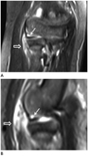
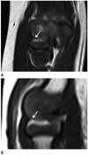
A 39-month-old child came to the emergency room of our hospital after he started to feel pain on the right forearm after he fell down on the trampoline. The child held the forearm in extension with full pronation and complained pain during supination and pronation on the physical examinations. There was no joint swelling, bruising, warmth, or erythema. No elbow fracture was identified on plain radiographs. Despite the absence of characteristic clinical history, nursemaid's reduction maneuver was performed several times under suspicion of pulled elbow based on clinical presentation and physical examinations. Subsequently, arm pain wasn't improved at all and an MRI was performed to assess other possible causes such as soft tissue, bone, and epiphysis injuries (Fig. 1). On the MR images, a displaced annular ligament in the right radiocapitellar joint combined with mild, edematous change of periarticular soft tissue was demonstrated without any abnormality of bony alignment of the arm. His right elbow was the splinted because severe pain was caused by repetition of unsuccessful manual reduction and the complete reduction could not be achieved, possibly due to periarticular soft tissue swelling and joint effusion. The pain had subsided and disappeared 3 weeks after the onset during the regular outpatient follow-up period. A follow-up MR imaging scan was taken 7 weeks after the symptoms subsided and spontaneous reduction of the ligament was found at this point (Fig. 2).
DISCUSSION
Pulled elbow is one of the most common traumatic elbow injuries in children under the age of 5 years. The infrequency of pulled elbow in children over the age of 5 is explained by relatively strong attachment of the annular ligament to the periosteum of the radial neck in Salter and Zaltz (3) anatomic investigations in 12 child cadavers.
Generally, the patient with pulled elbow presents an unwillingness to use the affected arm and the patient holds it limply at the side without deformity. With this characteristic clinical presentation, a typical history of abrupt longitudinal traction on the extended arm is sufficient for diagnosis in most cases.
However, there may be no characteristic history. This is often because the event was not witnessed. For this atypical case, radiographs should be preceed manual reduction since elbow fractures, such as supracondylar humerus fracture, might be misdiagnosed as pulled elbow (4, 5). If all types of elbow fractures are excluded, imaging studies such as MR may be helpful for diagnosis in atypical pulled elbow cases (6). Since the result of ultrasound for diagnosis depends most significantly on the operator, ultrasonography is difficult to standardize (7).
There are two standard methods of closed reductions for the treatment, which are supination and flexion or forced pronation of the affected arm (1, 8, 9, 10). According to a randomized clinical trial by Bek et al. (9), the hyperpronation maneuver was more efficient at the first reduction attempt, easier for physicians, and less painful for the patient even though final reduction rates were statistically similar. A click can be palpable, and sometimes audible, caused by the release of the entrapped annular ligament. Subsequently, the child usually shows full use of the affected arm a short period after the reduction maneuver. On the other hand, if there is no improvement of the symptoms even after the maneuver, immobilization with a sling or forearm cast for 1 week may be needed (8). And then the clinician should recheck the symptoms of the child.
Rarely, open reduction is required due to unsuccessful closed manipulation when the ligament escapes from the widest part of radial head (3, 8, 11). Delayed diagnosis also might prevent successful closed reduction (11).
Pulled elbow can recur within one week after treatment. Cases of early recurrence might be attributed to inadequate or incomplete reductions. One study showed a decrease in the recurrance rate after reduction and splint application with a flexed and supinated position (12).
Recently, Richardson et al. (6) reported an atypical pulled elbow case in which MRI was used to determine both the diagnosis of annular ligament entrapment and confirmation of successful reduction. When the patient came to visit the hospital with symptoms, clinicians tried to reduce the entrapped annular ligament under suspicion of pulled elbow. However the symptoms of the patients persisted. Then an MRI was performed and an entrapped annular ligament with normal bony alignment was confirmed. A more forceful reduction was attempted and successful with confirmed reduction of annular ligament by immediate follow-up MRI.
On the other hand, in the case of atypical pulled elbow in our hospital, we decided to immobilize the affected arm after failure of several trials of manual reduction and observe clinical symptoms through regular follow-up. The symptoms gradually improved and eventually disappeared within 3 weeks. The patient underwent MR imaging to confirm successful reduction of the entrapped annular ligament even though symptoms subsided. As a result, the follow-up MR imaging showed reduction of the entrapped annular ligament and no periarticular soft tissue edema.
To our knowledge, there has been no reported case in which spontaneous reduction was confirmed by follow-up MRI after subsequent immobilization of the affected arm with the splint due to unsuccessful manual reduction of atypical pulled elbow. The reason for unsuccessful reduction is not clear; it was possibly due to local soft tissue swelling or small hemorrhage in the region of the annular ligament (10).
As seen in our case of atypical pulled elbow (entrapped annular ligament) combined with only mild edematous change of periarticular soft tissue and no abnormality of bony alignment of the elbow, if the several trials of manual reduction are not successful, an excellent, stress-free treatment option might be to splint the elbow in a flexed and supinated position with subsequent follow-up.
In summary, spontaneous reduction of entrapped annular ligament by immobilization in an atypical pulled elbow patient with persistent symptoms after repeated trials of manual reduction was confirmed on MRI.
 XML Download