

 PDF
PDF ePub
ePub Citation
 Print
Print


INTRODUCTION
Coronary artery fistula (CAF) is a congenital anomaly defined as a direct precapillary connection between the coronary artery and other vascular structures. Its incidence is 0.1--0.2% in an adult population undergoing invasive angiography (1, 2). CAF usually terminates in the right ventricle, right atrium, and the pulmonary artery; CAF to the bronchial artery is very rare (3, 4, 5). The sinoatrial node (SAN) artery has various courses, and one important variant of this is the S-shaped SAN artery (6). Bronchial-to-SAN artery fistula has been mentioned in some previous reports, but a detailed description of its course on CT images has not been reported (4, 7). Herein we report a rare case of CAF to the bronchial artery from the S-shaped variant of the SAN artery, which was incidentally found on coronary angiography but its detailed course elucidated only after reviewing the coronary CT angiography (CCTA) images.
CASE REPORT
An 85-year-old male with nocturnal chest pain for three days visited our hospital. He had a history of diabetes mellitus and hypertension. No definite abnormal findings were noted on physical examination and electrocardiography. Creatine kinase MB, troponin I, and pro-brain natriuretic peptide levels were also within normal limit on laboratory examination. The chest radiograph was normal. However, due to persistent chest pain, the patient underwent coronary angiography with suspicion of coronary artery disease.
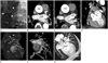
On selective coronary angiography through the left main coronary artery, a tortuous vessel arising from the proximal portion of left circumflex artery was revealed, which coursed upwards and branched into two smaller vessels. We suspected a CAF to the bronchial arteries. Another tiny vessel arising from the mid-portion of the tortuous vessel was also identified (Fig. 1A). No other abnormality was noted in the coronary arteries.
To accurately identify the course and the draining sites of the suspected CAF, electrocardiogram-gated CCTA was performed. On CCTA, a tortuous vessel was detected, arising from the proximal portion of the left circumflex artery and terminating in the right and the left bronchial arteries. On closer inspection, it was visible that this vessel ran posteriorly between the left atrial appendage and the ostium of the left pulmonary vein. The vessel divided into two branches at the posterosuperior portion of the left atrium, with the larger branch coursing upwards towards the bronchial arteries and the smaller branch running towards the junction of the superior vena cava and the right atrium (Fig. 1B-G). When the courses of the proximal half of the tortuous vessel and the smaller distal branch were put together, it was clear that they represented the course of the S-shaped SAN artery, an infrequent variant of the SAN artery.
The larger branch coursing upwards was the fistulous branch connecting the S-shaped SAN artery and the bronchial arteries. Thus, the coronary-to-bronchial fistula apparently arose from the middle of the S-shaped SAN artery near the posterosuperior aspect of the left atrium. No other abnormality was noted in the coronary arteries on CCTA.
However, because the CAF was not big enough to be clinically significant and the patient did not have any other abnormalities, the patient did not undergo intervention and was managed conservatively.
DISCUSSION
CAF is a congenital anomaly defined as a direct precapillary connection between the coronary artery and a cardiac chamber, a great vessel, or a systemic vessel. It is a rare condition with an incidence of 0.1--0.2% in an adult population undergoing diagnostic cardiac catheterization or coronary artery angiography (1, 2). It usually drains into the right ventricle (41%), right atrium (26%), and pulmonary artery (17%) (3). CAF has an infrequent connection to the bronchial arteries, with reported incidences of 0.55% (16/2922 cases) on angiography (4) and 0.61% (8/1300 cases) on CCTA.
The majority (75--81%) of coronary-to-bronchial artery fistula arises from the left circumflex artery, especially from the left atrial branch, and a small percentage arises from the right coronary artery (4, 5). Coronary-to-bronchial artery fistula is related to various cardiovascular and chronic pulmonary diseases including supravalvular aortic stenosis, Takayasu arteritis, pulmonary thromboembolism, bronchiectasis, and pulmonary tuberculosis. Most of the patients are known to be asymptomatic, although it can manifest as cardiac murmur, angina due to coronary steal phenomenon, infective endocarditis, rupture of an aneurysmal fistula, or hemoptysis. The SAN artery arises from either the right coronary artery (54.5%) or the left circumflex artery (40.6%) (6). The SAN artery is known to have various courses, and one important variant of this is the S-shaped SAN artery, which has an incidence of 14.6% in one study (6). The S-shaped SAN artery arises from the left circumflex artery and courses posteriorly between the left atrial appendage and the ostium of the left superior pulmonary vein and then anteriorly close to the anterior wall of the left atrium. Because of its proximity to the left atrial wall, potential vessel injury during cardiac intervention or surgery has been suggested.
Bronchial-to-SAN artery fistulas have been mentioned in some previous reports, but without detailed description of the course or accompanying CCTA images (4, 7). In fact, 9 out of 16 coronary-to-bronchial fistulas were from either the right or the left SAN artery in an angiographic study by Matsunaga et al. (4). However, as was in our case, it may be difficult to accurately identify the course and the anatomic relationship of the CAF and nearby structures with conventional angiography alone. The unknown vessel seen on the conventional angiography could be identified as the S-shaped SAN artery only after confirming the course of the vessel against adjacent structures on CCTA.
Because coronary-to-bronchial artery fistula can be a source of hemoptysis, especially in patients with bronchiectasis or pulmonary tuberculosis, embolization of the CAF via coronary artery is often done as treatment (7, 8). During the embolization of the coronary-to-bronchial artery fistula, if its origin from the SAN artery is not recognized, a disastrous embolization of the SAN artery could happen, resulting in the infarction of the SAN. Especially because the course of the S-shaped SAN artery is different from that of normally seen SAN artery, it could easily be mistaken as a tortuous, fistulous vessel. Therefore, intervention of CAF should be carefully done after inspection of the origin of the SAN artery. Confirming the accurate course of the CAF with CCTA may be necessary.
In summary, we report a rare case of coronary-to-bronchial artery fistula originating from an S-shaped SAN artery incidentally found on coronary angiography, in which CCTA was very helpful in accurately identifying the course and the origin of the artery.
 XML Download