

 PDF
PDF ePub
ePub Citation
 Print
Print


INTRODUCTION
Follicular thyroid carcinoma (FTC), which is the second common malignancy of the thyroid gland following papillary thyroid carcinoma (PTC), is usually diagnosed histologically by capsular and/or vascular invasion on permanent section. Meanwhile, it is more likely to metastasize to distant organs rather than to regional lymph nodes. The FTC may present with metastatic disease and the diagnosis can be made by histological examination of the metastatic disease (1). It is now well-known that the eggshell calcification of the thyroid nodule occurs in various benign and malignant diseases of the thyroid gland. Our review of the literature yielded four cases of FTC with eggshell calcification, with three of them presenting with distant metastasis and one with thyroid incidentaloma (2, 3).
We report here of a case of FTC in a 74-year-old male, with the interrupted eggshell calcification, presenting with renal and bony metastases along with the findings of primary and metastatic lesions at gray-scale, power Doppler ultrasonography (PD US), and CT scan.
CASE REPORT
A 74-year-old male presented with the poor oral intake and the pain, recently being aggravated, in the back and in both of the lower legs, which he had for 7 months following a traffic accident. The magnetic resonance imaging (MRI) of the lumbar spine, performed at another hospital 7 months prior, had revealed a suspicious metastatic lesion at the second lumbar vertebra and an enhancing mass in the lower pole of the right kidney. He had refused further examination, and had medicated at the local medical clinic for relief of pain. Otherwise, the physical examination was unremarkable. No remarkable abnormalities were noted on the routine laboratory tests, including thyroid function test.
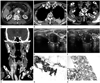
He underwent CT of the abdomen, chest, and neck by using SOMATOM Definition Flash scanner (Siemens Healthcare, Forchheim, Germany), for investigation of the unknown possible metastatic lesions of the second lumbar vertebra and the right kidney detected by MRI at another hospital. The CT of the abdomen and chest showed approximately 35 × 34 × 39 mm in size, exophytically grown, homogeneously enhancing mass at the lower pole of the right kidney, and homogeneously enhancing masses with bony destruction at the body, right pedicle of the second lumbar vertebra, and left fourth rib (Fig. 1A, B). The CT of the neck revealed a mass with interrupted eggshell calcification in the lower pole of the left thyroid lobe, which enhanced homogeneously following intravenous administration of contrast material (Fig. 1C, D). The sonographic examination, performed by using Acuson Sequoia 512 scanner (Siemens Medical Solutions, Mountain View, CA, USA) equipped with a 8- to 15-MHz linear array transducer, showed an approximately 17 × 20 × 23 mm-sized solid mass with interrupted eggshell calcification (Fig. 1E). The central and peripheral color signals were noted on PD US (Fig. 1F). He underwent sonographically guided fine-needle aspiration biopsy of the left thyroid mass with interrupted eggshell calcification, using 25-gauge needle attached to a 10-mL disposable syringe, which in turn was held by a reusable syringe holder. Three samples were obtained from the mass. We cautiously performed sonographically guided core-needle biopsy of the mass in the lower pole of the right kidney, by using a freehand technique with a 18-gauge needle (Bard Peripheral Technologies, Covington, GA, USA) and spring-loaded biopsy gun (Pro-Mag 2.2; Manan Medical Products, Northbrook, IL, USA). Two samples were obtained from the mass. The cytological and histological findings of the thyroid and renal masses were consistent with follicular neoplasm and metastatic follicular carcinoma, respectively (Fig. 1G, H). He was given dexamethasone and prednisolone for control of the back pain. He refused any further evaluation and management of FTC and metastatic diseases due to financial reasons and was lost to follow-up.
DISCUSSION
Our case presented diagnostic dilemma, because the primary lesion causing bony metastases to the vertebra and rib may be a renal mass or a thyroid mass with interrupted eggshell calcification. The genitourinary radiologist (SHK) interpreted this as a renal cell carcinoma with bony metastasis, while the head and neck radiologist (SKL) strongly suggested FTC with renal and bony metastases. Although the bony lesions of the second lumbar vertebra and the left fourth rib have not been histopathologically proven, we retrospectively speculated that they might be metastatic lesions from the primary FTC, because the patient had no known primary elsewhere and the enhancement patterns of the bony and renal lesions were nearly the same as that of the FTC with interrupted eggshell calcification.
The eggshell calcification on sonography can be defined as a curvilinear echogenic structure along the circumference of a nodule, and interruption of eggshell calcification as focal discontinuities of a curvilinear echogenic structure. To the best of our knowledge, two case reports concerning FTC with eggshell calcification have been in the literature (2, 3). Cheng et al. (2) reported the first case of FTC with eggshell calcification in a 63-year-old woman. They noticed a faint rim-like calcification in the right lower neck on the chest radiograph, which corresponded to a cold area of the right lower thyroid on Tc-99m pertechnetate scintigraph, and a focal osteolytic lesion involving frontal bone with epidural extension on the cranial CT. Ultrasound-guided fine-needle aspiration biopsy of the thyroid nodule and biopsy of the skull lesion proved to be follicular neoplasm and metastatic follicular carcinoma, respectively. However, they did not mention sonographic features of the thyroid mass.
We previously reported three cases of FTC with eggshell calcification (3). All three cases of FTC were spherical in shape and had interrupted eggshell calcification, and two had intranodular vacularity on PD US. Among them, two cases presented with distant metastasis.
Kim et al. (4) found that a hypoechoic halo and disruption of the eggshell calcification, which corresponded to the tumor invasion through (or over) eggshell calcification on pathologic examinations, may be more useful as sonographic predictors of PTC than known sonographic criteria including hypoechogenicity, irregular or microlobulated margins, and a taller-than-wide shape.
While the eggshell calcification may be lined inside of, within, or outside of the tumor capsule, the disruption of the eggshell calcification in the follicular neoplasm may be a useful finding that predicts capsular invasion. However, the patient refused to undergo surgical exploration and we could not obtain thyroidectomy specimen to verify sonographic-histologic correlation.
Distant metastasis is a major cause of thyroid cancer-specific mortality (5). Ito et al. (6) reported that distant metastasis at diagnosis is the strongest prognostic factor for cause-specific survival of the patients with widely invasive FTC, and the tumor size larger than 4 cm significantly affects the disease-free survival and cause-specific survival of the patients who did not show distant metastasis at diagnosis. The sizes of 3 cases, with FTC with interrupted eggshell calcification and distant metastasis, reported in the literature were 20 mm (2), 28.5 mm, and 10.3 mm (3) in the greatest diameter, and that of the present case was 23 mm. The size of a FTC with interrupted eggshell calcification that did not metastasize was 24.4 mm (3). It should be noted that the sizes of the masses of all FTC with interrupted eggshell calcification presenting with distant metastasis in the literature and present case were all less than 30 mm in the greatest diameter (range, 10.3-28.5 mm; average, 20.5 mm) (2, 3). Regarding the age of four patients with FTC with eggshell calcification presenting with distant metastasis, in the literature and present case, the average age was 71.8 years (range, 63-75 years) (2, 3). The age of a patient who had FTC with interrupted eggshell calcification, but without distant metastasis was 54 years (3). As such, interrupted eggshell calcification of FTC in elderly patients may be a poor prognostic sign, irrespective of the size of the tumor.
In summary, FTC should be included in the differential diagnosis of a thyroid mass with interrupted eggshell calcification. Furthermore, interrupted eggshell calcification itself in the elderly patients with FTC may frequently be associated with the distant metastasis. Thus, it may potentially represent poor prognostic sign rather than a favorable one. Further investigation with a large number of cases is anticipated, and intensive diagnostic investigations including surgical exploration may be warranted in elderly patients with cytologically suggested follicular neoplasm that has interrupted eggshell calcification.
 XML Download