

 PDF
PDF ePub
ePub Citation
 Print
Print


INTRODUCTION
Gardner syndrome is a rare autosomal dominant disorder characterized by colonic polyposis and bone and soft tissue manifestations including osteomas, and mesenchymal tumors of the skin and soft tissues (1, 2, 3). To our knowledge, unilateral chest wall anomaly has not been reported in patients with Gardner syndrome. We describe here a case of unilateral hypertrophy of the ribs and intercostals muscles occurring in a patient with Gardner syndrome.
CASE REPORT
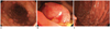
A 32-year-old man was admitted to the gastrointestinal department of our institution complaining of diarrhea and bloody stools. The patient had past histories of excision of lipomas from his left ear and back and hemorrhoidectomy. Familial history was not specific. A digital rectal examination revealed multiple polypoid masses. Insertion of a colonoscope as far as 40 cm above the anal verge showed extensive polypoid lesions along the entire colon (Fig. 1A). A 5 cm sized mass was observed 10 cm above the anal verge (Fig. 1B). Under the impression of familial adenomatous polyposis (FAP), the patient also underwent duodenoscopy, which showed numerous small polypoid lesions in the gastric body and multiple erythematous polypoid lesions in the duodenal bulb (Fig. 1C). Endoscopic biopsy of some of these lesions showed that they were adenomas with low grade dysplasia.
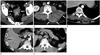
An abdominal and pelvic CT scan revealed an intraluminal polypoid mass with perirectal infiltrations in the upper rectum (Fig. 2A). The mass was regarded as the same lesion on previous colonoscopy (Fig. 1B), which was confirmed as being a tubular adenoma with low grade dysplasia following surgical excision. Axial postcontrast CT images showed soft tissue tumors with heterogenous density around the left paraspinal and quadratus lumborum muscles (Fig. 2B), the subcutaneous fat layer of the left thorax (Fig. 2C) and along the right ninth intercostal muscle (Fig. 2D), suggesting desmoids tumors. A repeat examination 377performed 19 months later showed no changes in these lesions. Precontrast CT showed two adrenal masses, of less than 10 Hounsfield units, which were diagnosed as adrenal adenomas (Fig. 2E).
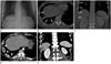
Based on the clinical, colonoscopic, and radiologic findings, the patient was diagnosed with Gardner syndrome. A preoperative chest radiograph (Fig. 3A) showed asymmetric hypertrophy of the right seventh to tenth ribs. Both lungs were clear but the volume of right lung showed slightly smaller than left one. Previous abdominal CT images showed increased size of the medullary portions of these lesions, but relatively normal thickness of the cortex (Fig. 3B, C). Intercostal muscles along the right seventh to tenth ribs were hypertrophied as compared with the contralateral ribs (Fig. 3D, E). The patient underwent prophylactic total colectomy with ileal pouch-anal anastomosis. The polypoid mass and numerous (more than 350) small polypoid lesions were histologically confirmed as tubular adenoma with low grade dysplasias.
DISCUSSION
Gardner syndrome is now recognized as a variant of FAP, since they share the same genetic alterations (4, 5). These disorders are linked to the adenomatous polyposis coli locus (APC gene), located at chromosomal band 5q21-q22. Approximately two thirds of all cases of Gardner syndrome are inherited, while one third are due to spontaneous mutation of the APC gene (4, 5). MYH (1p34.3-p32.1) is another gene associated with FAP (4). Symptoms are usually evident by the age 20 years, but they can present at any time between 2 months and 70 years. In general, the cutaneous and bone abnormalities develop approximately 10 years prior to polyposis (4, 6).
The gastrointestinal manifestations of Gardner syndrome include extensive adenomatous polyps in the colon, adenomatous polyps in the stomach and small intestine and periampullary carcinomas (1, 2, 4, 7). The colon is the most common site of involvement but other parts of the gastrointestinal tract may be involved (1). Most of the polyps are very small and difficult to visualize radiologically (4, 7). If left untreated, the potential for malignant transformation of colonic adenomatous polyps approaches 100% by the fourth decade of life (1, 4, 5). Prophylactic resection (total colectomy with construction of an ileoanal pouch) should be performed before age 25 years, ideally between ages 16 and 20 years (4, 7). Since duodenal or jejunal adenomatous polyps and their malignant transformations may occur after colectomy, patients should be carefully monitored and larger adenomas endoscopically removed (7).
More than half of patients with FAP or Gardner syndrome have osseous involvement (7). The most common bone abnormalities observed in patients with Gardner syndrome are osteomas consisting of dense osseous lesions originating on the bone surface (6). Almost all parts of the skeletal system may be involved. Rib abnormalities have been reported, including wavy and localized cortical thickening or osteomas of several ribs (2, 6, 8). Except for osteomas, few patients show other types of osseous manifestations, including osteosarcoma and intraosseous pilomatricoma (5, 7). The recognition of osteomas is important because they frequently precede the appearance of intestinal polyposis and may be an early sign of this disorder (6).
The most common soft tissue lesions of Gardner syndrome are epidermoid cysts on the face, scalp, and extremities (4, 5, 7). Other soft tissue tumors and tumor-like lesions include fibromas, neurofibromas, lipomas, leiomyomas, and pigmented skin lesions (1, 9). Desmoid tumors appear in 3.5-5.7% of patients and they can appear at any time (4). Common locations are the incision sites, the abdominal cavity and the retroperitoneum. These tumors are considered among the most troublesome manifestations of Gardner syndrome, since they may cause life threatening complications and are usually resistant to treatment (3).
To our knowledge, unilateral chest wall anomaly has never been reported in patients with Gardner syndrome. There has been no report about a reduced long volume in patients with Gardner syndrome. This rare abnormality is possibly related to Gardner syndrome and also can be an incidental finding. The recognition of unilateral hypertrophy of the ribs can be an early sign of Gardner syndrome as well as osteomas. The possible hypothesis for unilateral chest wall anomaly is compensatory overgrowth of chest wall due to a small lung volume. However the degree of reduced lung volume is minimal and the patient had no pulmonary symptoms. Inversely hypertrophied chest wall might restrict the growth of right lung.
In conclusion, we have described a patient with unilateral hypertrophy of ribs and intercostal muscles in a patient with Gardner syndrome. Careful attention is necessary to recognize the variable and minute bone abnormalities in patients with Gardner syndrome.
 XML Download