

 PDF
PDF ePub
ePub Citation
 Print
Print


Abstract
Purpose
To evaluate the accuracy of abdominal ultrasonography (US), and the incidence, size and types of lesions, easily missed on abdominal US in health screening.
Materials and Methods
We retrospectively enrolled 311 physical checkup patients (male-to-female ratio, 156:155; mean age, 53.1 years), who underwent screening abdominal US and abdominal computed tomography (CT) on same day from July, 2011 to October, 2013. Per-organ and per-lesion analyses and verification of size and location of renal and hepatic lesions which were frequently missed on abdominal US were performed.
Results
Overall, 209 additional lesions were found on abdominal CT in 140 physical checkup patients and the missing rate of the lesion was 45%. Renal lesions were most common (105 lesions), followed by hepatic lesions (91 lesions). In renal and hepatic lesions which were missed on abdominal US, most (93.4%, 93.5%) lesions were less than 1.5 cm in longitudinal diameter. Most of missed renal lesions were located in mid portion of left kidney (24.7%). Most of the missed hepatic lesions on US were located in the hepatic dome (segment 7, 8, 2) (66%).
Figures and Tables
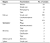
Fig. 1
A 65-year-old male with adrenal adenoma in right adrenal gland (arrow), that was not found on abdominal ultrasonography, though it could be detected on contrast enhanced abdominal CT.
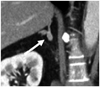
Fig. 2
A 42-year-old female with renal stone.
A. Ultrasonography shows no abnormal finding in right kidney.
B. There is a tiny right renal stone (arrow), that was not found on ultrasonography, in lower pole on contrast enhanced abdominal CT.
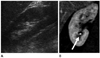
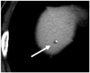
Fig. 3
A 50-year-old male with tiny hepatic cyst with peripheral calcification (arrow) in S8 of the liver, that was not found on abdominal ultrasonography.

References
1. Bennett GL, Krinsky GA, Abitbol RJ, Kim SY, Theise ND, Teperman LW. Sonographic detection of hepatocellular carcinoma and dysplastic nodules in cirrhosis: correlation of pretransplantation sonography and liver explant pathology in 200 patients. AJR Am J Roentgenol. 2002; 179:75–80.
2. Kudo M, Okanoue T. Japan Society of Hepatology. Management of hepatocellular carcinoma in Japan: consensus-based clinical practice manual proposed by the Japan Society of Hepatology. Oncology. 2007; 72:Suppl 1. 2–15.
3. Bruix J, Sherman M, Llovet JM, Beaugrand M, Lencioni R, Burroughs AK, et al. Clinical management of hepatocellular carcinoma. Conclusions of the Barcelona-2000 EASL conference. European Association for the Study of the Liver. J Hepatol. 2001; 35:421–430.
4. Bruix J, Sherman M. Practice Guidelines Committee, American Association for the Study of Liver Diseases. Management of hepatocellular carcinoma. Hepatology. 2005; 42:1208–1236.
5. Park JW, Choi JY, Seo KS, Jeong JW, Seong JS, Kim JW, et al. Practice guidelines for management of hepatocellular carcinoma 2009. Seoul: Jingihoek;2009. p. 3–7.
6. Bolondi L, Sofia S, Siringo S, Gaiani S, Casali A, Zironi G, et al. Surveillance programme of cirrhotic patients for early diagnosis and treatment of hepatocellular carcinoma: a cost effectiveness analysis. Gut. 2001; 48:251–259.
7. Zhang B, Yang B. Combined alpha fetoprotein testing and ultrasonography as a screening test for primary liver cancer. J Med Screen. 1999; 6:108–110.
8. Lee CW, Choi JI, Kim MJ, Lee JS, Jung DC, Lee CY, et al. Added value of screening low dose computed tomography of the chest for the evaluation of abdominal solid organs by ultrasound in physical check-up patients. J Korean Soc Ultrasound Med. 2010; 29:97–104.
 XML Download