

 PDF
PDF ePub
ePub Citation
 Print
Print


Abstract
Unilateral pulmonary vein atresia is a rare congenital anomaly. Its symptoms begin to manifest in childhood and a broad spectrum of clinical severity has been described, ranging from asymptomatic, recurrent pulmonary infection, severe hemoptysis, to death. Only a few adult cases with this condition, with no or mild symptoms, have been reported. Pulmonary angiography has been typically used for definite diagnosis. However, pulmonary angiography may be replaced with the current developing multidetector CT. This report presents an adult case with mild symptoms, diagnosed by multidetector CT.
Figures and Tables
Fig. 1
Rt. pulmonary vein atresia in 16-year-old man. Chest radiograph shows a small right hemithorax and diminished vessel size in the right lung with slight mediastinal displacement to the right.
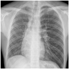
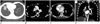
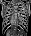
Fig. 2
Rt. pulmonary vein atresia in 16-year-old man.
A. Axial CT scan (lung window) shows small right hemithorax with mild interlobular septal thickening in right lower lobe and right middle lobe.
B. Contrast-enhanced axial CT scan (mediastinal window) shows the small right pulmonary artery and normal left pulmonary artery.
C, D. Axial and coronal CT scan shows smooth margin of the left atrium with no pulmonary vein (thin arrows) and numerous dilated vessels seen on paraesophageal area, representing bronchial collaterals (thick arrow).
References
1. Heyneman LE, Nolan RL, Harrison JK, McAdams HP. Congenital unilateral pulmonary vein atresia: radiologic findings in three adult patients. AJR Am J Roentgenol. 2001; 177:681–685.
2. Pourmoghadam KK, Moore JW, Khan M, Geary EM, Madan N, Wolfson BJ, et al. Congenital unilateral pulmonary venous atresia: definitive diagnosis and treatment. Pediatr Cardiol. 2003; 24:73–79.
3. Kim Y, Yoo IR, Ahn MI, Han DH. Asymptomatic adults with isolated, unilateral right pulmonary vein atresia: multidetector CT findings. Br J Radiol. 2011; 84:e109–e113.
4. Savaş Bozbaş Ş, Varan B, Akçay Ş. Right pulmonary venous atresia: a case report and review of literature. Tuberk Toraks. 2012; 60:254–257.
5. Dixit R, Kumar J, Chowdhury V, Rajeshwari K, Sethi GR. Case report: isolated unilateral pulmonary vein atresia diagnosed on 128-slice multidetector CT. Indian J Radiol Imaging. 2011; 21:253–256.
6. Mataciunas M, Gumbiene L, Cibiras S, Tarutis V, Tamosiunas AE. CT angiography of mildly symptomatic, isolated, unilateral right pulmonary vein atresia. Pediatr Radiol. 2009; 39:1087–1090.
7. Cao M, Cai H, Ding J, Zhuang Y, Wang Z. Bronchial varices in congenital unilateral pulmonary vein atresia. Am J Respir Crit Care Med. 2013; 187:1267–1268.
8. Harris KM, Lloyd DC, Morrissey B, Adams H. The computed tomographic appearances in pulmonary artery atresia. Clin Radiol. 1992; 45:382–386.
9. Oguz B, Haliloglu M, Karcaaltincaba M. Paediatric multidetector CT angiography: spectrum of congenital thoracic vascular anomalies. Br J Radiol. 2007; 80:376–383.
10. Gasparetto TD, Daltro P, Marchiori E. Imaging findings of an asymptomatic child with pulmonary vein atresia. Pediatr Radiol. 2010; 40:1458–1459.
 XML Download