

 PDF
PDF ePub
ePub Citation
 Print
Print


Abstract
Idiopathic pleuroparenchymal fibroelastosis (IPPFE) is a recently described, very rare type of fibrotic interstitial lung disease predominantly involving subpleural areas of both upper lungs. IPPFE has distinctive radiologic and pathologic features: progressive subpleural opacity with fibrotic changes, predominantly in upper lungs, and dense elastic component on histology. We experienced one case of surgically confirmed IPPFE, with progression of radiologic findings on the serial CT examinations. We herein report the characteristic radiologic features of IPPFE with pathologic and clinical manifestations.
Figures and Tables
Fig. 1
A 57-year-old female who showed fibrotic interstitial disease predominantly involves both apical lungs.
A. Chest radiograph shows bilateral irregular opacities with volume loss and hilar elevation in the subpleural area of both upper lungs.
B, C. CT shows bilateral subpleural irregular opacities, pleural thickening (long arrows) and traction bronchiolectasis (short arrows) in both upper lobes on lung window setting (B) and mediastinal setting (C).
D. CT shows subpleural reticular opacities (long arrows) with mild traction bronchiectasis (short arrow) in both lower lobes.
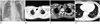
Fig. 2
Coronal reformation CT images shows progressive subpleural irregular opacities and traction bronchiectasis with volume loss in both upper lungs, and less progressive subpleural reticular opacities with volume loss in both lower lungs than upper lungs; 8 years ago (A), 2 years ago (B), present (C).
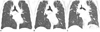
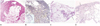
Fig. 3
Histopathologic features suggesting idiopathic pleuroparenchymal fibroelastosis, which revealed by surgical lung resection.
A. Right upper lobe lung shows dense fibrosis along the subpleura (arrow). The fibrosis extends into away from the pleura and shows perilobular distribution with alveolar septum (haematoxylin and eosin, × 2).
B. Dense elastic fibers (arrows) are irregularly scattered throughout the fibrosis on Elastic Van Geison stain (elastic Van Gieson staining, × 20).
C. Right lower lobe lung parenchyma shows typical microscopic features of idiopathic pulmonary fibrosis including subplerual fibrosis, patchy distribution, temporal heterogeneity and interstitial fibroblastic foci with microscopic honeycomb change (arrow) (haematoxylin and eosin, × 2).
D. The fibrotic area of subpleura shows small amount of deposition of elastic fiber (arrow) which is as same as normal lung pleura and parenchyma (elastic Van Gieson staining, × 20).
References
1. Travis WD, Costabel U, Hansell DM, King TE Jr, Lynch DA, Nicholson AG, et al. An official American Thoracic Society/European Respiratory Society statement: update of the international multidisciplinary classification of the idiopathic interstitial pneumonias. Am J Respir Crit Care Med. 2013; 188:733–748.
2. Reddy TL, Tominaga M, Hansell DM, von der Thusen J, Rassl D, Parfrey H, et al. Pleuroparenchymal fibroelastosis: a spectrum of histopathological and imaging phenotypes. Eur Respir J. 2012; 40:377–385.
3. Frankel SK, Cool CD, Lynch DA, Brown KK. Idiopathic pleuroparenchymal fibroelastosis: description of a novel clinicopathologic entity. Chest. 2004; 126:2007–2013.
4. Watanabe K, Nagata N, Kitasato Y, Wakamatsu K, Nabeshima K, Harada T, et al. Rapid decrease in forced vital capacity in patients with idiopathic pulmonary upper lobe fibrosis. Respir Investig. 2012; 50:88–97.
5. Piciucchi S, Tomassetti S, Casoni G, Sverzellati N, Carloni A, Dubini A, et al. High resolution CT and histological findings in idiopathic pleuroparenchymal fibroelastosis: features and differential diagnosis. Respir Res. 2011; 12:111.
6. Becker CD, Gil J, Padilla ML. Idiopathic pleuroparenchymal fibroelastosis: an unrecognized or misdiagnosed entity? Mod Pathol. 2008; 21:784–787.
7. Amitani R, Niimi A, Kuse F. [Idiopathic pulmonary upper lobe fibrosis (IPUF)]. Kokyu. 1992; 11:693–699.
8. Repo UK, Kentala E, Koistinen J, Lehtipuu AL, Miettinen A, Pyrhönen S, et al. Pulmonary apical fibrocystic disease. A serologic study. Eur J Respir Dis. 1981; 62:46–45.
9. Shiota S, Shimizu K, Suzuki M, Nakaya Y, Sakamoto K, Iwase A, et al. [Seven cases of marked pulmonary fibrosis in the upper lobe]. Nihon Kokyuki Gakkai Zasshi. 1999; 37:87–96.
10. Ryerson CJ, Collard HR. Update on the diagnosis and classification of ILD. Curr Opin Pulm Med. 2013; 19:453–459.
 XML Download