

 PDF
PDF ePub
ePub Citation
 Print
Print


INTRODUCTION
Neurofibromatosis type 1 (NF-1) is a autosomal dominant inherited condition. The diagnostic criteria for this disease include development of café au lait spots, plexiform neruofibroma, axillary or groin freckling, optic glioma, Lisch nodule formation, distinctive bony lesions, and a first-degree relative with NF-1. Patients with NF-1 may have additional features such as intracranial tumors, macrocephaly, pheochromocytoma, neurologic impairment, vascular stenosis, or aneurysms. Vascular involvement of NF-1 is very rare and may present as an aneurysm, obstruction, or stenosis. Vasculopathy associated with NF-1 may result in hemothorax, a condition that can result in fatal complications or sudden death (1).
CASE REPORT
A 34-year-old man developed sudden onset left pleuritic pain and dyspnea 5 days before admission to the emergency room. He had a palpable, nonpulsatile swelling on the left side of his neck. The patient also had multiple café au lait spots and polyposis all over his body. His mother had a history of multiple cutaneous neurofibromas. The patient had sinus tachycardia and hypophonesis on the left hemithorax. His blood pressure was 110/70 mm Hg with a heart rate of 104 beats/min, respiratory rate of 24 breaths/min, and oxygen saturation of 97%. His hematocrit value was 25% with a hemoglobin level of 8.8 g/dL.
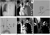
A large degree of left pleural effusion and atelectasis of the left lung were observed on the chest radiograph, and the trachea had shifted to the right thorax (Fig. 1A). Thoracic computed tomography (CT) revealed a massive left hemothorax and passive atelectasis. In addition, a pseudoaneurysm (maximum diameter 3.8 cm, 2.4 cm in neck size) was detected in the left proximal vertebral artery with a perianeurysmal hematoma. The left subclavian artery was compressed by the aneurysm with a hematoma (Fig. 1B). The patient was referred to the interventional unit for emergent embolization of the aneurysm. Selective vertebral angiography showed a pseudoaneurysm 3.8 cm in size originating from the left proximal vertebral artery and 80% narrowing of the left mid-subclavian artery (Fig. 1C). The patient underwent embolization of the efferent vertebral artery with microplex 7EA coils (5 mm/15 cm, Microvention Terumo Medical Products, Somerset, NJ, USA) to occlude proximal and distal blood flow of the vertebral aneurysm. However, coil embolization of the afferent vertebral artery failed as the afferent segment was adjacent to the origin of the left subclavian artery. Additionally, an uncovered stent 1EA (10/60 mm, SNG, Seoul, Korea) was inserted into the compressed left subclavian artery. After coil embolization and stent deployment, patency of left subclavian artery and occlusion of distal portion of left vertebral artery are confirmed by thoracic aortography (Fig. 1D). The patient was subsequently transferred to the operating room for an exploratory thoracotomy and evacuation of the hematoma. A 4 L hematoma was removed during the operation.
The patient was stable after surgery. However, he experienced numbness and pain in the left forearm. Thoracic CT was repeated 13 days after embolization and surgery. An in-stent occlusion within the left subclavian artery was observed (Fig. 1E). A bypass graft from the left carotid artery to the left subclavian artery was performed, and the hematoma around the left vertebral artery was removed (Fig. 1F). Postoperative electromusculography for persistent pain and paresthesia in the left forearm indicated a left brachial plexus injury at the trunk level. The patient was transferred to the rehabilitation department and his condition improved with medication, physical therapy, and occupational therapy.
DISCUSSION
A spontaneous hemothorax due to an intrathoracic arterial rupture in cases of NF-1 is rare. The incidence of vasculopathy in patients with NF-1 is only 3.6% (2) including individuals with aneurysms and a stenosis. Aneurysms may rupture and result in a fatality. Rupture of aneurysms in the subclavian (3), intercostal (4), bronchial (5), and vertebral arteries (6) have been reported in patients with NF-1. Salyer and Salyer (7) suggested that intimal thickening accompanying NF-1 vasculopathy is the result of Schwann cell proliferation within the arteries. This demonstrates a pathogenic relationship between vasculopathy and neurofibromas that characterize NF-1. An aneurysm in a patient with NF-1 is characterized by fibrous intimal thickening, irregular loss of smooth muscle media, and elastic membrane fragmentation. These features often cause weakening of the supportive connective tissue and adjacent muscle coat in the wall of the artery, which could cause rupture of the arterial wall (6). A more recent study showed that smooth muscle cells rather than Schwann cells proliferate in cases of NF-1 vasculopathy (7).
Diagnostic modalities for patients with NF-1 and vasculopathy include CT or magnetic resonance angiography, chest X-ray, chest ultrasound, and thoracentesis. Among these methods, CT angiography is the best method for assessing vascular lesions and the selection of treatment by endovascular intervention (8, 9). CT can show detailed anatomy, and is less invasive. We believe that postoperative CT angiography was important for our case not only because it allowed us to evaluate complications and the prognosis, but it was also a non-invasive procedure and alleviated patient pain during the evaluation. We were also able to detected stent occlusion and obliteration of the pseudoaneurysm by follow-up CT angiography.
Therapeutic maneuvers for treating a massive hemothorax due to an intrathoracic aneurysm include surgical and non-surgical techniques. As this disease can be fatal, emergency operations such as thoracotomy with excision of lesion have been performed. However, some recent reports demonstrated that non-surgical methods such as endovascular intervention or stent-graft placement are preferable for treating hemothorax arising from an aneurysm rupture in cases of NF-1 (3, 4, 9). In addition, a good prognosis of patients undergoing these procedures has been described (2, 10). Endovascular coil embolization is a useful, less invasive, and a safe method for controlling arterial hemorrhage in cases that do not require preservation of arterial flow. In our patient, we performed endovascular coil embolization and also inserted a stent into the adjacent stenotic lesion. NF-1 vasculopathy may cause further hemodynamic and neurologic complications. Accordingly, the effects of vasculopathy and resulting secondary consequences should be considered.
In conclusion, vasculopathy associated with NF-1 is rare condition. However, ruptured aneurysms can cause a fatal hemothorax and secondary complications such as blood flow disturbances. Imaging techniques to determine the extent of the lesion, detect complications, and plan treatment methods including endovascular intervention or surgery should be carefully performed.
 XML Download