

 PDF
PDF ePub
ePub Citation
 Print
Print


Abstract
Purpose
To evaluate the effectiveness of percutaneous biliary stone removal as a primary treatment in cases with difficulties to use an endoscopy.
Materials and Methods
From March 2004 to May 2011, 17 patients who underwent primary percutaneous biliary stone removal (Group 1) and 34 case-matched patients who underwent primary endoscopic biliary stone removal were selected (Group 2). The inclusion criteria were as follows: patients who had 1) ≥ 15 mm bile duct stones, 2) intrahepatic bile duct stones, 3) bile duct stones with a history of previous gastrointestinal bypass surgery. In the present study were analyzed the success rates, the length of postprocedural hospital stay, the change of Amylase/Lipase values and complications post procedure. Statistical analysis was performed using paired t-test and unpaired t-test.
Figures and Tables
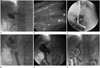 | Fig. 1A 87-year-old woman with distal common bile duct stones.
A. Cholangiogram shows a large impacted stones (55, 18 mm in each diameter) in the distal common bile duct.
B, C. On endoscopic examination, one stone was broken using by mechanical lithotripsy. But lithotripsy for large proximal stone was failed.
D. The ampullary sphincter was dilated using a 10 mm-sized balloon catheter.
E. The stone was captured and fragmented using 7 Fr stone basket. The fragmented stones were completely extracted.
F. Follow-up cholangiogram shows no demonstrable residual stone in the distal common bile duct.
|
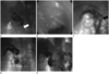 | Fig. 2A 77-year-old woman with distal common bile duct stone. She underwent total gastrectomy with esophago-jejunostomy due to gastric cancer.
A. Cholangiogram shows an impacted stones in the distal common bile duct (white arrow).
B. On endoscopic examination, the ampullary orifice could not be found due to previous operation.
C. The ampullary sphincter was dilated using a 10 mm-sized balloon catheter (stone diameter: 13 mm, black arrow).
D. The stone was captured and pulled using 7 Fr stone basket. The fragmented stone was completely extracted.
E. Follow-up cholangiogram shows no demonstrable residual stone in the distal common bile duct.
|


References
1. Lim JH. Oriental cholangiohepatitis: pathologic, clinical, and radiologic features. AJR Am J Roentgenol. 1991; 157:1–8.
2. Kim SH, Sohn CH, Kim YH. Percutaneous lithotripsy for removing difficult bile duct stones using endoscopy. J Korean Radiol Soc. 2008; 58:229–236.
3. Kawai K, Akasaka Y, Murakami K, Tada M, Koli Y. Endoscopic sphincterotomy of the ampulla of Vater. Gastrointest Endosc. 1974; 20:148–151.
4. Vaira D, D'Anna L, Ainley C, Dowsett J, Williams S, Baillie J, et al. Endoscopic sphincterotomy in 1000 consecutive patients. Lancet. 1989; 2:431–434.
5. Clouse ME, Stokes KR, Lee RG, Falchuk KR. Bile duct stones: percutaneous transhepatic removal. Radiology. 1986; 160:525–529.
6. Neuhaus H. Intrahepatic stones: the percutaneous approach. Can J Gastroenterol. 1999; 13:467–472.
7. Park YS, Kim JH, Choi YW, Lee TH, Hwang CM, Cho YJ, et al. Percutaneous treatment of extrahepatic bile duct stones assisted by balloon sphincteroplasty and occlusion balloon. Korean J Radiol. 2005; 6:235–240.
8. Seon HG, Kwon CI, Yoon SP, Yoo KH, Ok CS, Kim WH, et al. PTPBD for managing extrahepatic bile duct stones in patients with failed or contraindicated ERCP. Korean J Med. 2012; 83:65–74.
9. Ozcan N, Kahriman G, Mavili E. Percutaneous transhepatic removal of bile duct stones: results of 261 patients. Cardiovasc Intervent Radiol. 2012; 35:890–897.
10. van der Velden JJ, Berger MY, Bonjer HJ, Brakel K, Laméris JS. Percutaneous treatment of bile duct stones in patients treated unsuccessfully with endoscopic retrograde procedures. Gastrointest Endosc. 2000; 51(4 Pt 1):418–422.
11. Cho DH, Park GT, Oh JE, Chung CW, Yoo GJ, Kim SR, et al. [A single institution's experience of endoscopic retrograde cholangiopancreaticography in the eldery patients: outcomes, safety and complications]. Korean J Gastroenterol. 2011; 58:88–92.
12. Mondet AF. [Technic of blood extraction of calculi in residual lithasis of the choledochus]. Bol Trab Soc Cir B Aires. 1962; 46:278–290.
13. Burhenne HJ. Garland lecture. Percutaneous extraction of retained biliary tract stones: 661 patients. AJR Am J Roentgenol. 1980; 134:889–898.
14. Burhenne HJ. Nonoperative retained biliary tract stone extraction. A new roentgenologic technique. Am J Roentgenol Radium Ther Nucl Med. 1973; 117:388–339.
15. Gil S, de la Iglesia P, Verdú JF, de España F, Arenas J, Irurzun J. Effectiveness and safety of balloon dilation of the papilla and the use of an occlusion balloon for clearance of bile duct calculi. AJR Am J Roentgenol. 2000; 174:1455–1460.
16. Miller BM, Kozarek RA, Ryan JA Jr, Ball TJ, Traverso LW. Surgical versus endoscopic management of common bile duct stones. Ann Surg. 1988; 207:135–141.
17. Neoptolemos JP, Carr-Locke DL, Fraser I, Fossard DP. The management of common bile duct calculi by endoscopic sphincterotomy in patients with gallbladders in situ. Br J Surg. 1984; 71:69–71.
18. Siegel JH, Safrany L, Ben-Zvi JS, Pullano WE, Cooperman A, Stenzel M, et al. Duodenoscopic sphincterotomy in patients with gallbladders in situ: report of a series of 1272 patients. Am J Gastroenterol. 1988; 83:1255–1258.
19. Ozcan N, Erdogan N, Baskol M. Common bile duct stones detected after cholecystectomy: advancement into the duodenum via the percutaneous route. Cardiovasc Intervent Radiol. 2003; 26:150–153.
20. Meranze SG, Stein EJ, Burke DR, Hartz WH, McLean GK. Removal of retained common bile duct stones with angiographic occlusion balloons. AJR Am J Roentgenol. 1986; 146:383–385.
21. Ilgit ET, Gürel K, Onal B. Percutaneous management of bile duct stones. Eur J Radiol. 2002; 43:237–245.
22. Stokes KR, Clouse ME. Biliary duct stones: percutaneous transhepatic removal. Cardiovasc Intervent Radiol. 1990; 13:240–244.
23. Maydeo A, Bhandari S. Balloon sphincteroplasty for removing difficult bile duct stones. Endoscopy. 2007; 39:958–961.
24. Ersoz G, Tekesin O, Ozutemiz AO, Gunsar F. Biliary sphincterotomy plus dilation with a large balloon for bile duct stones that are difficult to extract. Gastrointest Endosc. 2003; 57:156–159.
25. García-García L, Lanciego C. Percutaneous treatment of biliary stones: sphincteroplasty and occlusion balloon for the clearance of bile duct calculi. AJR Am J Roentgenol. 2004; 182:663–670.
 XML Download