

 PDF
PDF ePub
ePub Citation
 Print
Print


INTRODUCTION
In 1995, Evans (1) first described a desmoplastic fibroblastoma (DF), a fibrous soft tissue tumor comprising spindle shaped cells to stellate fibroblastic cells sparsely distributed in a dense fibrous background. This tumor, an alternative also called collagenous fibroma (2), was clinically and morphologically distinct, as well as completely benign in previously reported series. Details of magnetic resonance imaging (MRI) findings for such a tumor have been described for only a few cases (3, 4).
In this article, we present the case of a DF of the foot and we will discuss about the MRI findings of this tumor.
CASE REPORT
A 44-year-old man presented a palpable mass in the plantar aspect of the left foot. He was otherwise feeling well with no reports in the medical history or a previous foot trauma. The laboratory samples results were normal.
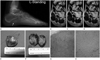
The foot radiography showed an about 3.2 × 2.0 cm sized slightly dense soft tissue mass at the plantar aspect of his left foot (Fig. 1A).
In a MRI, a 2 × 2.8 × 2.1 cm sized ovoid shaped mass lesion in the medial plantar aspect of the left foot was shown. On T1-weighted images [repetition time (TR) = 436 ms, echo time (TE) = 12 ms] the mass was well circumscribed, homogenous and had slightly low signal intensity (SI) compared with adjacent muscles (Fig. 1B). The mass was located beneath the abductor hallucis muscles and medial to the flexor digitorum brevis muscle. The lesion had a mass effect upon the adjacent muscles. On the T2-weighted images (TR = 3179 ms, TE = 60 ms), the mass had mixed SI as high SI within low SI (Fig. 1C). The post contrast T1-weighted images (TR = 548 ms, TE = 12 ms) did not show any enhancement (Fig. 1D).
An excisional biopsy was performed which revealed a firm encapsulated mass (Fig. 1E, F). This mass was totally removed. As a pathological result the tumor had well circumscribed margins and was composed of a proliferation of spindle and stellate fibroblast-like cells sparsely distributed in a collagenous matrix. The final diagnosis was desmoplastic fibroblastoma (collagenous fibroma) (Fig. 1G, H).
DISCUSSION
The desmoplastic fibroblastoma (collagenous fibroma) are more present in adult male patients with a mean age 50 years at diagnosis. Most of the DFs arise from the extremities, shoulder girdle, posterior neck, upper back or the abdominal wall. The tumors usually are variable in size (1.0-20.0 cm). Clinically, the DF displays clinical typically as a painless, mobile, firm and well-circumscribed subcutaneous mass which grows slowly with an enlargement duration of more than 6 months. In general, the tumors appear oval, disc shaped or fusiform and have a firm homogenous pearl-gray colored consistency on cut-section (5).
Microscopic most of the DFs show low a cellularity and consist of spindle and stellate shaped fibroblastic cells without atypical or hyperchromatic nuclei (3). Also, in case of doubts, immunohistochemistry aids the correct diagnosis. Especially, the S-100 protein, CD34 and EMA are useful markers for an exclusion of neurofibroma, solitary fibrous tumor and perineuroma (5).
There are only a few previous case reports of DF with MRI features present. Beggs et al. (6) described a single case of synovial DF of the hip joint. Magnetic resonance imaging showed a mass with a minimally high signal intensity on T2-weighted images. Gadolinium enhanced images were not performed.
In our case, the tumor had a smooth well defined margin, inhomogeneous low signal intensity in the T2-weighted images. The area showed slightly high SI on T2-weighted images which corresponded to the hypercellular area within the lesion that consisted of a tumor with loose collagen fibers. The low T2 signal intensity is attributed to the thick abundant collagen background (5).
The most of the soft tissue masses have a high signal intensity on T2-weighted images. Soft-tissue masses with a low SI on T2-weighted images include neurofibroma, cicatricial fibroma, malignant fibrous histiocytoma, aggressive fibromatosis and calcified masses (myositis ossificans, extraskeletal osteosarcoma or chondrosarcoma and synovial sarcoma) (7).
Abundant collagen and marked hypocellularity in a soft-tissue tumor in the absence of calcification result in a decreased signal on the T2-weighted pulse sequence.
Other malignant neoplasms, such as malignant fibrous histiocytoma and fibrosarcoma may also have a low SI within the lesions. Because these malignant neoplasms infiltrate to the surrounding tissue and have usually a necrotic/cystic change, the majority of these tumors can be differentiated from DF due to a MRI (8).
The desmoid tumor is the most important differential diagnosis which should be considered (8, 9). The desmoid tumor is histological more cellular, vascular, and infiltrative at its periphery than the DF. On imaging studies, a desmoid tumor is not as clear circumscribed as a DF. The desmoid tumor may also have larger areas which show a very high SI on T2-weighted images and represents more cellularity than the DF.
The treatment of DF is surgical resection. In our case neither local recurrence nor metastasis occurred after resection which also differed from the high local recurrence rate of a desmoid tumor.
In summary, we described a patient with a DF arising from the foot. This disease should be considered when low SI-areas with poor contrast enhancement in a mass with well-defined margins are seen on both T1- and T2-weighted images.
 XML Download