

 PDF
PDF ePub
ePub Citation
 Print
Print


INTRODUCTION
Carpal tunnel syndrome (CTS) is a common disorder caused by compression of the median nerve within the carpal tunnel. The disorder is most frequently idiopathic. However, the space occupying lesion or systemic disease could contribute to CTS (1). Extraskeletal chondroma is a rare, benign, cartilaginous, soft tissue tumor, usually occurring in the hands and the feet (2, 3, 4). There are few reports in the literature on carpal tunnel syndrome caused by extraskeletal chondroma (5). We present a case of carpal tunnel syndrome, as a result of an extraskeletal chondroma arising within the carpal tunnel, with descriptions on the radiological and pathological findings of the mass. Authors also discuss the differential diagnosis of the calcified space occupying lesions that may occur in carpal tunnel.
CASE REPORT
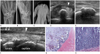
A 64-year-old female patient presented with a five-year history of a progressively painful and tingling sensation in her hand. A clinical examination showed wasting in her thenar eminence and altered sensation in the median nerve distribution. Tinel's and Phalen's tests of the median nerve at the carpal tunnel level were both positive. There was no specific past medical or family history. Radiographs of the wrist, including carpal tunnel view, demonstrated a well-defined, oval, homogeneously calcified lesion within the carpal tunnel, just ventral to the capitate (Fig. 1A-C). A ultrasound (US) study demonstrated a well-defined, echogenic lesion with posterior acoustic shadowing, measuring 15 × 8 × 5 mm in size. The calcified mass was surrounded by the flexor tendons, and was located in the floor of carpal tunnel (Fig. 1D, E). The mass was not movable with finger motion in US. The vascularity of the lesion was not detected on Doppler US scan. The median nerve flattened at the carpal tunnel, with a hypoechoic swollen appearance at the proximal side of the tunnel (Fig. 1D, F). In the views of imaging, our differential diagnosis of the lesion included hydroxyapatite deposition disease (HADD) and extraarticular synovial chondromatosis. The patient underwent carpal tunnel release and total excision of the lesion, with complete decompression of the median nerve. There was no connection between the lesion and the carpal bones. Histologically, the excised tumor was encapsulated by a thick fibrous capsule and showed nodular configuration (Fig. 1G). Microscopically, the tumor consisted of mature chondrocytes, which were irregularly divided by interlobular fibrous and myxoid septa. Calcium deposits surrounded benign looking chondrocytes and/or intercellular stroma. A mixed hypocellular zone, with myxoid background in the center and hypercellular zone in the periphery, was seen (Fig. 1G, H). These histopathologic features were consistent with the benign catilagenous tumor. The tumor was diagnosed as an extraskeletal calcified chondroma. Six weeks following the surgery, the patient had no residual painful tingling sensation in her fingers.
DISCUSSION
CTS are most frequently idiopathic. However, some space occupying lesions or systemic diseases might cause CTS. Systemic diseases may include diabetes, amyloid deposits, and hypothyroidism. The space occupying the lesion may include inflammation, trauma, tumors, or anatomical anomalies. Among these, tenosynovitis of the flexor tendons and ganglion are the most often encountered (1). Both increase in content or decrease in size of the carpal canal may elevate the pressure within the carpal tunnel and cause symptoms. In patients with idiopathic CTS, symptoms are typically bilateral and particularly prevalent in middle-aged woman. When patients present with an atypical feature, the possibility of another cause of CTS, such as space occupying lesion, should be considered (1). Extraskeletal chondroma is an unusual and benign soft tissue mass. It is small and usually is a well-defined nodule of cartilage that is unattached to the bones. The mass constitutes approximately 1.5% of all benign soft tissue tumors. It mainly affects the patients of 30 to 60 years of age. But the mass may be encountered over a wide age range (2, 3, 4). This benign tumor is usually discovered as a solitary lesion in the soft tissue of the hand and the foot. Clinically, the mass usually grows slowly, occasionally causing pain or tenderness. Lesions are typically well demarcated and lobulated, rarely exceeding 2 cm in greater dimension, firm and rubbery on palpation, and often mobile. The extraskeletal chondroma is not reported to metastasize or to undergo malignant transformation, and the rate of local recurrence is reported to be 15-20% (3). Local excision appears to be the treatment of choice (3). The histopathological appearance of extraskeletal chondroma is variable, ranging from an immature pattern dominated by chondroblasts to a mature form with chondrocytes. It contains matrix of hyaline cartilage, fibrosis, calcification or ossification, or myxoid content. A granuloma-like reaction may be seen within the stroma. Radiologically, calcification is observed in 33-70% of the cases, and may have curvilinear, punctuate, mixed pattern, and dystrophic or focal dense pattern. Extrinsic bone erosion or sclerosis can be associated (2-4). There are scant literatures on US findings of extraskeletal chondroma. Bianchi et al. (6) reported that extraskeletal chondroma is revealed as a well-defined hypoechoic mass with good posterior acoustic enhancement. Park et al. (7) reported a case of soft tissue chondroma represented by a well-demarcated, hypervascular, hypoechoic mass with posterior acoustic enhancement on US. There were no radiological or histological calcifications reported in masses in the previous studies. In this case, the mass radiographically showed the dense calcification. It appeared as a well-defined, hypovascular, echogenic lesion with posterior acoustic shadowing on US (Fig. 1D-G). These discrepant US findings seemed to be the results of the various degrees of calcification in the mass from case to case. So, conventional and Doppler US features of extraskeletal chondroma can be varied, and can depend on the amount of calcification of the mass and the response of the surrounding tissue. Based on the literatures, the tumors have non-specific findings on MR imaging, and have shown high signal intensity on T2-weighted images (T2WI), intermediate signal intensity on T1-weighted images (T1WI), and hypointensity of corresponding areas both on T1WI and T2WI, when significant calcification is seen (2). When extraskeletal chondroma occurs in carpal tunnel, especially in the presence of calcifications as in our case, it should be differentiated from other benign soft tissue lesions, including periosteal chondroma, extraarticular synovial chondromatosis, crystal deposition diseases such as HADD, calcium pyrophosphate deposition disease, gout, and tumoral calcinosis (8). Typically, periosteal chondroma is juxtacortical, and is attached to the bone from within and beneath periosteum. Radiologically, it presents as a sharply margined bone surface tumor, often with calcification and mineralization of the chondroid matrix, which are classically associated with saucerization of the underlying bone. Extraarticular synovial chondromatosis is usually characterized by the multiple, small, metaplastic cartilaginous or osseo-cartilaginous nodules, attached to the synovium of the tendon sheath or the extraarticular bursa, whereas extraskeletal chondroma tends to form a well-defined solitary mass (9). Unlike extraskeletal chondroma, the calcification tends to be amorphous, nodular, or multilobulated, and have relatively poor margins in crystal deposition diseases (10). Malignant soft tissue tumors showing calcification also should be distinguished. They may include synovial sarcorma, extraskeletal myxoid chondrosarcoma, and extraskeletal osteosarcoma. Synovial sarcoma usually shows indistinct margin, less frequently has calcifications often at the periphery, and is spotty (4). Extraskeletal chondrosarcoma and extraskeletal osteosarcoma are extremely rare in the hands and the feet. They tend to be larger in deep soft tissues, and they have more irregular calcifications or ossification than that of extraskeletal chondroma (8).
In summary, extraskeletal chondroma may rarely occur in carpal tunnel and result in CTS. The radiographic and the US features of extraskeletal chondroma seem to be non-specific and are various, particularly depending on the amount of calcification of the tumor. But, when a small, slowly growing, well-demarcated, calcified soft tissue mass is detected, particularly in the distal extremities including the carpal tunnel, extraskeletal chondroma should be included in the differential diagnosis of the lesion.
 XML Download