

 PDF
PDF ePub
ePub Citation
 Print
Print


INTRODUCTION
Azygos vein has many variations in their venous tributaries. Among them, the absence of the azygos vein is a very rare form of the congenital venous anatomic variation, and only a few cases have been reported in the literature up to recently (1, 2, 3). We report the radiologic findings of the congenital absence of the azygos vein with hemiazygos vein, which drained into the left superior vena cava via superior intercostal vein (SICV).
CASE REPORT
A 65-year-old man diagnosed as rectal cancer visited our colorectal cancer clinic. He did not show any chest symptoms.
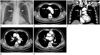
He underwent preoperative chest posteroanterior radiograph and postoperative contrast-enhanced chest computed tomography (CT) to monitor metastasis in the thorax. On the chest radiograph, azygos arch shadow was not visible in its usual location, and there was an aortic nipple, a focal bulge adjacent to the aortic arch, representing the left SICV (Fig. 1A). The chest CT showed no azygos vein at its usual location and the left SVC was present (Fig. 1B-D). Hemiazygos vein was slightly dilated and drained into the left SVC via prominent left SICV (Fig. 1D, E).
DISCUSSION
Because of the complexity of its developmental stages, the cardinal vein system could undergo a variety number of congenital anomalies during the embryonic period (4). While in the developmental period, the cardinal system consists of anterior and posterior cardinal veins. By the eighth week, right and left anterior cardinal veins become connected by an anastomotic duct, which later become the superior vena cava on right side (from right anterior cardinal vein and right common cardinal vein) and left brachiocephalic vein on left side (from left anterior cardinal vein) (3, 4). However, when the left common cardinal vein fails to be obliterated, it becomes the left SVC (4).
The second most important system of the great thoracic veins is the subcardinal veins that form the azygos and the hemiazygos veins (4). Azygos vein is corresponded by the right subcardinal vein and hemiazygos vein by the left subcardinal vein. The connection of the right and left subcardinal veins usually forms at the level of the sixth or seventh thoracic vertebra (5). The left subcardinal vein undergoes obliteration cranial to the anastomotic site, or it may persist as the accessory hemiazygos vein. The accessory hemiazygos vein is connected to the left SICV medial to the distal aortic arch (6). The left SICV is developed from the embryonic posterior cardinal veins. The left SICV then courses anteriorly beside the aortic arch to meet the left brachiocephalic vein posteriorly (6).
A persistent left SVC is considered to be the most common anomalous systemic vein-to-cardiac connection which is seen in 0.3-0.5% of general population (7). Up to 90% of the people with persistent left SVC present with the right SVC as well. About 65% of the people with persistent left SVC have no left brachiocephalic vein or atrophic one. About 20% of them show left superior intercostal vein forming a communication between hemiazygos vein and the left SVC, producing a left hemiazygos arch like the current case (7).
There have been several case reports about the radiologic findings of the absence of azygos vein (1, 2, 3, 5, 6, 7). All of these cases showed no azygos vein at the normal anatomic position and the "aortic nipple" in the dependent drainage side (i.e., left SVC, left innominate vein) on the chest radiograph. Chest CT showed the "aortic nipple" and the agenesis of the azygos vein. It also showed some cases with the right SVC only (1) or the left SVC only (2, 3, 5), and the patients with bilateral SVC (6, 7) like in this case.
Absence of the azygos vein is a very rare congenital venous anomaly, but we should take this anomaly into account when the chest radiograph fails to show azygos arch shadow on its usual location and shows the "aortic nipple" on the lateral side of aortic arch. The chest CT is the best way to confirm the agenesis of the azygos vein and the associated thoracic venous anomalies based on the chest radiograph.
 XML Download