

 PDF
PDF ePub
ePub Citation
 Print
Print


INTRODUCTION
The most cases of anomalous origins of the vertebral artery (VA) are incidental findings because these variations are clinically asymptomatic (1). Recently, technological advances in CT and MR angiography imaging allow a better delineation of anatomical variations as well as vascular pathologies in the major intracranial and cervical arteries. Although an anomalous origin of the right vertebral artery (RVA) is rare, the identification of this variation is important for the performance of endovascular procedures or cardiothoracic surgeries (2). In this paper, a rare case of an aberrant RVA originated from the aortic arch distal to the left subclavian artery on CT angiography will be presented, the relevant literatures on this variation will be reviewed and their embryologic development and clinical implications will be discussed also.
CASE REPORT
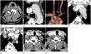
A 34-year-old man visited after a stabbing injury of the neck the emergency department with a hemiparesis right. The patient was alert awake and had stable vital signs on arrival. An initial neurologic examination revealed decreased muscle strength of the right extremities as 1/5 lower extremity power and 2/5 right upper extremity power. Following laboratory findings were within normal ranges: complete blood count, serum electrolytes, liver function test, serum creatinine and routine urinalysis. The patient underwent a contrast-enhanced CT angiography for the evaluation of combined vascular injuries. The CT scan revealed a linear foreign body with low density (chopstick) in the right central spinal canal at C4-5 level without an associated vascular injury (Fig. 1A). Incidentally, it was also detected an anomalous origin of the RVA arising from the aortic arch distal to the left subclavian artery with Kummerell's diverticulum (Fig. 1B, C). The prevertebral segment of the RVA was located in the retroesophageal and retrotracheal areas (Fig. 1D, E). Also it had an aberrant entrance to the C7 transverse foramen while the left VA showed a normal entrance to the C6 transverse foramen (Fig. 1F, G).
DISCUSSION
Although the VA is classically the first branch of the ipsilateral subclavian artery, multiple anomalous origins of the VA have been reported in the literature (1). With a reported prevalence of 2.4-5.8% in a large series of autopsies is the left VA arising from the aortic arch between the left common carotid artery and left subclavian artery the most common variation of a VA origin (2, 3, 4). An aberrant RVA is an extremely rare anomaly and may confuse an aberrant right subclavian artery by its retroesophageal course, especially when it is stenotic (5, 6). This variant is divided into three categories: first, those directly originating from the aorta; second, those originating from the carotid or brachiocephalic arteries; and third, those with duplicated origin (2, 4). Embryologically, the VA is formed by the development of postcostal longitudinal anastomosis which links the cervical intersegmental arteries. The intersegmental arteries eventually obliterated with an exception of the seventh, which becomes the proximal subclavian artery and includes the VA point of origin in adults (3, 7, 8). An aberrant RVA originating from the aortic arch distal to the left subclavian artery is compatible with the persistence of the proximal dorsal aorta on the right side and with the segment regression of the right dorsal aorta between the sixth and seventh intersegmental arteries (3, 7, 8). The RVA is the only branch to stay connected to the persistent proximal dorsal aorta and arises distal to the left subclavian artery if the right subclavian artery originates from the seventh intersegmental artery normally (1, 3).
With only 11 cases published in the literature previously the aberrant RVA as the last branch of the aortic arch is a very rare variation (6, 9, 10). In the most cases described in the literature, there were no clinical signs or symptoms related with this variant (2, 3, 4). In the present case the aberrant RVA showed an anomalous course of the prevertebral segment located in the retroesophageal and retrotracheal areas with an aberrant entrance to the C7 transverse foramen. In contrast to previous reports, the patient presented a neurological deficit associated with a spinal cord injury after a stabbing accident.
A detailed knowledge about aberrations of the VA origin is potentially important to avoid an inadvertent vascular injury and an associated ischemic event of the brainstem during the endovascular procedures or a cardiothoracic surgery (1, 2, 4, 5). Therefore, the possibility of such a variant must be considered if a VA cannot be found in the usual position.
In conclusion, we provide a rare case of an aberrant RVA as the last branch of the posterior aspect of the aortic arch in a patient with a stabbing injury. With this report it will be suggested that an awareness of anomalous origins of the VA and its embryologic mechanism may be helpful for an identification of this variant on CT or MR angiographies in clinical practices.
 XML Download