

 PDF
PDF ePub
ePub Citation
 Print
Print


INTRODUCTION
Leiomyosarcoma is a malignant mesenchymal tumor which is composed of cells showing smooth muscle differentiations. Primary leiomyosarcoma of the gallbladder is a rare malignancy with poor prognosis. The imaging findings of gallbladder leiomyosarcoma are rarely reported. We present the ultrasonography (US), computed tomography (CT), and positron emission tomography-computed tomography (PET-CT) imaging findings of a primary leiomyosarcoma on the gallbladder in an 82-year-old male.
CASE REPORT
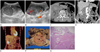
An 82-year-old male was referred to our institution from a local clinic due to an incidentally detected large gallbladder mass. The patient complained of vague right upper abdominal discomforts. Laboratory findings revealed mild leukocytosis and elevated liver enzymes (hemoglobin, 11.3 g/dL; hematocrit, 33.2%; red blood cell count, 3.60 × 10³/µL; white blood cell count, 11.9 × 10³/µL; neutrophil, 78%; platelet count, 225 × 103/µL; alanine transaminase, 292 IU/L; aspartate transaminase, 314 IU/L; total bilirubin, 1.81 mg/dL; direct bilirubin, 0.69 mg/dL). Tumor markers were within the normal limits (alpha-fetoprotein, 1.52 IU/mL; cancer antigen 19-9, 1.82 IU/mL). Abdominal ultrasound showed a large polypoid intraluminal protruding mass, displaying heterogeneous echogenicity and relatively well-defined margins with displaced thick hypoechoic overlying mucosa. Intratumoral vascularity was remarkably increased on color Doppler ultrasonography. In the lumen of the gallbladder, there were mottled echogenic foci with posterior acoustic shadows suggesting gallstones, and also heterogeneous hyperechoic materials suggesting hematoma besides the mass. Extraluminal invasion was not shown (Fig. 1A, B). Contrasting material enhanced abdominal CT scans demonstrated a large polypoid mass with broad base arising from the body of the gallbladder, which is approximately 5.5 cm in diameter, protruding into the lumen. The mass showed smooth border forming obtuse angles with the nearby gallbladder wall, lobulated margin, heterogeneous enhancement with extensive necrosis, and hemorrhage. Overlying mucosa was intact and thick, indicating a relatively clear demarcation line with the mass. Intense contrast enhancement with dilated tortuous vessels originating from cystic artery was detected. The gallbladder was distended and its wall is irregularly thickened. It also contained numerous gallstones and hemorrhages. Subserosal infiltration, hepatic metastasis, and metastatic lymphadenopathy were not seen in the scans (Fig. 1C, D). PET-CT revealed the large mass as a remarkably hypermetabolic lesion (standardized uptake value = 8.3) (Fig. 1E).
A provisional diagnosis of gallbladder carcinoma was made. Radical cholecystectomy with hepatic wedge resection was performed via laparotomy. Gross specimens demonstrated a large polypoid mass with a broad base arising from the body of the gallbladder. It contained numerous necrotic and hemorrhagic foci (Fig. 1F). Histopathology revealed that tumor cells are located below the mucosal and muscle layer, and spindle cells with fascicular patterns are found in the tumor (Fig. 1G).
The immunohistochemistry showed positive immunoreactivity for vimentin and smooth muscle actin, but negative immunoreactivity for pancytokeratin, epithelial membrane antigen, S-100 protein, and c-kit. The confirmed diagnosis of gallbladder leiomyosarcoma was made.
DISCUSSION
Gallbladder sarcomas are rare, with less than 200 cases reported (1). Gallbladder leiomyosarcomas are extremely rare and the estimated frequency is 1.4 per 1000 gallbladder malignancies (2). Our search through PubMed revealed that this case is the 22nd in this series (1, 3-6).
Gallbladder leiomyosarcoma occurs more frequently in females than in males during their sixth to seventh decades of life. Patients with gallbladder leiomyosarcoma are presented with abdominal pain, fever, jaundice, palpable mass, hepatomegaly, weight loss, or anorexia. Biochemically, they showed elevated alkaline phosphatase and bilirubin, or leukocytosis resembling those of acute cholecystitis or gallbladder carcinoma (3, 5). Gallstones and chronic inflammatory changes have been suggested as the predisposing factors in pathogenesis of gallbladder leiomyosarcoma, although no proofs exist to date (7).
Despite the advances in radiology, it is still hard to differentiate leiomyosarcoma from usual adenocarcinoma or other unusual malignant tumors of the gallbladder prior to the surgeries. Although not all the available literatures on gallbladder leiomyosarcoma have described the gross pathological or radiological features, descriptions of a polypoid mass protruding into the lumen and a dilated gallbladder with irregularly thickened wall have been widely published (3-5, 8, 9). The CT and US images in our case also showed similar radiological and gross pathological features with the aforementioned cases mentioned, and the radiological features overlapped with those of gallbladder adenocarcinoma and other neoplastic polyps. However, in this case, the mass showed smooth borders forming obtuse angles with the nearby gallbladder wall, and the overlying thick mucosal layer was displaced, suggesting submucosal lesions, which are unlike the normal findings for gallbladder adenocarcinoma.
In our case, differential diagnosis included leiomyosarcoma, fibrosarcoma, carcinosarcoma, and gastrointestinal stromal tumors. The radiologic differential diagnosis of solid polypoid gallbladder tumor is very difficult. However, if the tumor is located at the gallbladder wall and the overlying mucosal layer is displaced by the tumor, it is suggestive of submucosal tumors of the gallbladder, such as gastrointestinal stromal tumor or leiomyosarcoma.
Only histological examinations can differentiate leiomyosarcoma from other sarcoma or mesenchymal tumors. When the cells show positive immunoreactivity for Vimentin and smooth muscle actin, but negative immunoreactivity for cytokeratins AE1/A3, CD117, CD34, and S-100 protein on immunohistochemistry, the leiomyosarcoma can be differentiated from other sarcomas or mesenchymal tumors such as gastrointestinal stromal tumors (1, 10).
The treatment of choice is radical cholecystectomy with regional lymph node dissections as usual malignant tumors of the gallbladder. More radical procedures such as adjacent organ resection, major hepatectomy, pancreaticoduodenectomy, and portal vein resection can also be performed (5, 7).
The prognosis is considered very poor for patients with gallbladder leiomyosarcoma due to the highly malignant nature associated with rapid progression and early metastases. Most patients die within 5 years despite their radical procedures (6).
In conclusion, primary gallbladder leiomyosarcomas are extremely rare, and it is hard to preoperatively differentiate primary gallbladder leiomyosarcoma from other malignant tumors of the gallbladder such as adenocarcinoma. However, the leiomyosarcoma can be included in the differential diagnosis of primary gallbladder malignancy, particularly when radiological images show a hypervascular polypoid mass protruding into the gallbladder lumen and overlying thick mucosal layer.
 XML Download