

 PDF
PDF ePub
ePub Citation
 Print
Print


INTRODUCTION
Diffuse cavernous hemangioma (DCH) of the large bowel is a rare cause of gastrointestinal bleeding. DCH is often seen in young patients. While colonic localization is very uncommon, the rectosigmoid is the most common site in the gastrointestinal tract. To date, approximately 100 cases of DCH of the rectosigmoid colon have been reported in the literature (1). Although the tumor is uncommon, it is very important for it to be detected by radiologists because its accurate diagnosis is crucial for avoiding a biopsy, as a biopsy could cause severe hemorrhage (2). We report a case of DCH occurring in the transverse colon and present the CT and MR imaging, and pathologic findings.
CASE REPORT
A 41-year-old man was admitted due to bleeding after a colonoscopic biopsy. The patient had had no symptoms or problems prior to this presentation. At that time, he underwent colonoscopy for screening, and bleeding could not be controlled after the biopsy, so the patient was brought to our hospital.
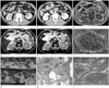
There were no specific findings on physical examination, including skin lesions, and routine laboratory test results were normal. CT revealed a diffuse circumferential wall thickening of the right transverse colon and adjacent hepatic flexure with several small nodular dense calcifications, and a hemostatic clip deployed by a previous colonoscopy (Fig. 1A). The thickened wall of the colon showed an undulated inner margin and outer contour, as well as heterogeneous enhancement with highly enhanced small nodules (Fig. 1B). Also, multiple highly enhanced small nodules and several small nodular dense calcifications were diffusely disseminated in the pericolic fat adjacent to the lesion. On MR imaging, there was a circumferential wall thickening of the right transverse colon and hepatic flexure which revealed slightly low signal intensity on the T1-weighted image (Fig. 1C), high signal intensity on the T2-weighted image (T2-WI) (Fig. 1D), and high signal intensity on the heavily T2-WI, compared to that of the mesenteric fat (Fig. 1E).
The impression was an unusual hemangioma of the transverse colon, and colonoscopy was performed. Colonoscopy showed a reddish, hyperemic mucosa with a bluish varix-like protruding lesion, which was thought to be a submucosal vessel dilatation. The patient underwent segmental resection of the transverse colon. On gross pathological examination, the transverse colonic mucosa exhibited a huge, ill-defined, markedly congested, bluish purple, discrete to mulberry-like, conglomerated submucosal tumefaction (Fig. 1F). The cut sections revealed numerous, blood-filled, sponge-like, microcystic spaces, scattered at the submucosa, the muscularis propria and the pericolic adipose tissue (Fig. 1G). Histopathologic examination under a low power view revealed multiple closely apposed multilocular blood-filled thin walled vascular channels, sharing a thin fibrocollagenous common wall, at the submucosa and the muscularis propria (Fig. 1H). On the high power view, multilocular cavernous vascular channels were lined by bland-looking, single endothelial cells, often containing inraluminal, fresh or organizing, fibrin thrombi and a minimal focus of dystrophic calcification (Fig. 1I).
DISCUSSION
Hemangioma of the large bowel can be of the capillary or cavernous type, and ranges from well-circumscribed polypoidal masses to diffusely infiltrative lesions. The cavernous type is more common (75-80%) and is characterized by large thin-walled vessels with smooth muscle fibers and connective tissue stroma primarily in the submucosal location. On the other hand, capillary hemangioma is formed by a network of closely packed well-circumscribed vessels and is often asymptomatic (3). DCH of the large bowel occurs frequently in children and young adults (age range, 5-25 years). The most common site affected (50-70%) is the rectosigmoid colon. Since patients usually give a long history of recurrent painless rectal bleeding, they frequently have some degree of anemia.
Radiographs of the abdomen provide an important diagnostic clue by revealing clusters of phleboliths, which are seen in 26-50% of adult patients, although they are uncommon in young children (4). CT findings include a concentrically thickened rectosigmoid wall with heterogeneously enhancing perirectal soft tissue and tubular structures. An accurate diagnosis of DCH can be made on the basis of these findings in conjunction with calcified phleboliths in the colonic wall and perirectal soft tissue. Since colon cancer typically has an intermediate signal intensity between the high signal intensity of the fat tissue and the low signal intensity of the muscular layer on T2-WI, DCH of the colon is easily differentiated from colon cancer on MR (5).
Colonoscopy is essential in the evaluation of colon hemangioma. Hemangiomas appear as submucosal projections, ranging from deep blue to dull red ("plum" color) (6). In the stable patient with no active bleeding, a complete survey of the colon and the upper gastrointestinal tract is important in excluding synchronous lesions. Biopsy may cause severe hemorrhage and is usually contraindicated (2).
Tung et al. (7) have shown that the signal intensity characteristics depend on the relative composition of the vascular spaces and connective tissue within the lesion as well as the presence of thrombosis, calcification, hemorrhage, or fibrosis. Similarly, delayed enhancement of the hepatic hemangioma is probably caused by a longer retention of contrast material in large intravascular spaces, resulting from slow flow, puddling, and partial thrombosis in these sites (8). In our case, the pathology showed multiple intraluminal fresh to organized thrombosis in vascular channels, but other findings, such as calcification, hemorrhage, and fibrosis, were not significant. Furthermore, the contrast between the lesion and pericolic fat was more remarkable on heavily T2-WI than on conventional T2-WI, which allowed for the easy detection of the lesion.
In conclusion, our case showed progressive nodular high enhancement and phleboliths on CT, and high signal intensity on MR T2-WI and heavily T2-WI. The clinical features of DCH are nonspecific and performing a biopsy for diagnosis is fraught with the risk of bleeding. Therefore, noninvasive imaging assumes a great significance in the diagnosis and management of this clinical condition. CT and MR imaging findings, especially hyperintensity on T2-WI, and progressive nodular high enhancement on contrast-enhanced images, are specific for DCH of the large bowel.
 XML Download