

 PDF
PDF ePub
ePub Citation
 Print
Print


INTRODUCTION
Lipomas are the most common benign tumors of the small intestine, generally solitary and asymptomatic (1). However, intestinal lipomatosis characterized by diffuse, multiple, and circumscribed lipomas in the intestine is a rare disease (2, 3). Furthermore, complications of intestinal lipomatosis including intussusception, volvulus, and ulceration are extremely rare (3). Here we present a case of lipomatosis in the jejunum with volvulus presenting as an intestinal obstruction.
CASE REPORT
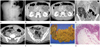
A 50-year-old male was admitted with severe colicky epigastric pain for a day. His personal history was unremarkable. On physical examination, the abdomen was soft and the epigastrium showed mild tenderness. The results of laboratory studies including routine blood tests, liver function tests, and urine analysis were normal. His upright conventional abdominal radiograph revealed dilatation of the small bowel with multiple air-fluid levels in the left abdomen, which indicated a mechanical ileus (Fig. 1A). Contrast-enhanced computed tomography (CT) demonstrated twisted mesenteric vessels in a swirling pattern and dilatation of the proximal small bowel in the left upper quadrant (Fig. 1B, C). In addition, multiple, homogeneous, fat-attenuating, intraluminal masses (-85 and -100 Hounsfield unit) with smooth margins were found in the small bowel loop in the right lower quadrant and pelvic cavity (Fig. 1D-F). No demonstrable enhancing focus was noticed within the fatty masses. These imaging features were highly suggestive of intestinal lipomatosis. As there was a combined small bowel volvulus on CT, laparotomy was performed.
During laparotomy, the intestinal loop was found to be markedly distorted and dilated, which is consistent with the CT findings of the volvulus. The point of twist was a proximal jejunal loop with a counterclockwise rotation. There was no evidence of vascular compromise. Adhesiolysis was required to decompress the volvulus. Moreover, multiple intraluminal masses were palpable within the proximal jejunum 40 cm from the Treitz ligament and were about 40 cm in length. Multiple diverticulae were also identified in this segment. At least 60 cm of the affected segment of the jejunum was resected.
Gross pathological examination revealed numerous intraluminal polypoid masses (over one hundred) measuring up to 6 cm in diameter and 40 cm in length (Fig. 1G). The cut surfaces of the masses were yellowish and fatty. Microscopically, the intraluminal masses were composed of mature adipocytes in the submucosa (Fig. 1H). Numerous diverticulae, up to 4 cm in diameter, were present in the resected small intestine.
The patient's postoperative period was uneventful and a follow-up examination after 6 months revealed no recurrence of lipomas.
DISCUSSION
Most lipomas in the small intestine are single. Intestinal lipomatosis is a rare condition that was first reported by Hellstrom in 1906 (3). Intestinal lipomatosis is characterized by the presence of numerous circumscribed lipomas in the intestine (3). Pathologically, the proliferation of adipose cells can be confined to the submucosa or may extend to the serosa or mesentery. The muscularis propria is usually not affected (4).
Intestinal lipomatosis shows no gender predominance and a wide range of age distribution from 20 to 88 years (mean 47 years) (3). Most cases are asymptomatic. Clinical manifestations are related to the presence of complications such as intussusception, volvulus, or ulceration (5).
Intestinal obstruction caused by intussusceptions or volvulus can result in recurrent abdominal pain. Some patients complain of bleeding or anemia due to inflammation, necrosis, or ulceration of the mucosa underlying the condition (3, 5).
Relatively higher prevalence of diverticulosis among patients with lipomatosis has been reported in previous studies (3, 5-7). Although the muscularis propria is usually unaffected, lipomatosis in the submucosal layer can play a contributory role in the development of diverticulosis by weakening the bowel wall (3, 7). Furthermore, lipomatosis may cause intermittent or incomplete obstruction and increase intraluminal pressure, which encourages the development of pulsion diverticulae (7). In our case, multiple diverticulae were identified in both macroscopic and microscopic examinations. During the retrospective review of the patient's CT images, we recognized small air-filled outpouchings of the affected small bowel wall, which were suggestive of diverticulae (Fig. 1D-F).
A few cases of volvulus with intestinal lipomatosis have been diagnosed (3, 5, 8). In our patient, CT revealed typical findings of intestinal lipomatosis of the jejunum in the right lower quadrant and pelvic cavity, which are not the usual locations of the jejunum. It is probably due to the counterclockwise rotation of the small bowel and its mesentery associated with volvulus. The abnormal weight distribution of the bowel may also be a factor for its occurrence (3).
CT can be one of the essential modalities to diagnose the primary lesion and its complications such as intussuception or volvulus. Moreover, CT is particularly useful for differentiating fat from other soft tissues such that fatty masses of the small intestine can be easily detected. Through CT, multiple homogeneous masses with a density equal to that of normal fat in the small intestine are characteristic findings of intestinal lipomatosis (1, 9). It is important for radiologists to differentiate the malignant nature of fat-containing tumors such as liposarcoma. Liposarcoma can be distinguished by its inhomogeneous appearance as it contains connective tissue myxoid elements or lymphocytic infiltration (10). Fortunately, sarcomatous changes in intestinal lipomatosis have never been reported (3, 10).
There is no uniform consensus regarding the treatment of patients with intestinal lipomatosis (6). Specific treatment is not required for asymptomatic patients (1). Subsequent hemorrhage or intussuception can be complicated; in such cases, cure is achieved after surgical resection of the affected area (3). In the case of volvulus, only derotation may be performed (3).
In conclusion, we report a rare case of intestinal lipomatosis with volvulus substantiated by characteristic CT findings. Although its incidence is rare, it is important for radiologists to be aware of intestinal lipomatosis since it can be easily and accurately diagnosed using CT findings.
 XML Download