

 PDF
PDF ePub
ePub Citation
 Print
Print


INTRODUCTION
Oncocytoma of the ocular adnexa is a rare benign tumor that consists of transformed glandular epithelial cells with abundant eosinophilic granular cytoplasm (1).
Since Radnót (2) introduced the concept of ocular oncocytoma in 1941, > 60 cases of oncocytoma of the ocular adnexa have been reported (3). Oncocytomas account for 3.5% of all lacrimal sac neoplasms (4) and 3-8% of all biopsied caruncular masses (5). Oncocytoma of the ocular adnexa is reportedly prevalent in elderly women (6). Most cases of orbital oncocytoma arise from the caruncle, followed by the lacrimal sac (3). Among them, malignant oncocytoma of the orbit is especially rare.
Here we report a case of a 76-year-old man who was diagnosed with a malignant oncocytoma.
CASE REPORT
A 76-year-old man presented with a 10-year history of a left orbital mass. The patient noticed the mass 10 years prior but did not seek treatment since it caused neither pain nor discomfort.
Facial computed tomography (CT) scans revealed a 4.0 × 3.0 × 3.3 cm relatively well-circumscribed, homogeneously enhancing mass in the left orbit. The mass showed high attenuation [77 Hounsfield unit (HU)] without calcification on a pre-contrast CT scan and good enhancement (140 HU) after contrast media administration (Fig. 1). It was located in the medial and inferior portions of the intraconal, conal, and extraconal spaces abutting the left eyeball.
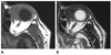
Before the CT scans, he underwent brain magnetic resonance (MR) imaging due to two episodes of transient ischemic attacks. The intervals were 8 months between the initial and follow-up brain MR images and 2 months between the CT scans and the follow-up brain MR images. The size and signal intensity of the mass did not change significantly between either the initial and follow-up brain MR images or the CT scans and follow-up brain MR images. This mass showed isosignal intensity on a T1-weighted image (WI) and low signal intensity on a T2WI (Fig. 2). No restricted diffusion was observed on diffusion weighted imaging (DWI) and apparent diffusion coefficient (ADC) mapping.
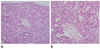
An incisional biopsy was performed under local anesthesia. Histopathological examination revealed the proliferation of atypical oncocytic neoplastic cells with stromal invasion, which is suggestive of malignant oncocytoma (Fig. 3).
Because of the malignant features of the lesion, exenteration was recommended, but the patient refused surgery and chose radiation therapy instead. There was no evidence of metastatic lesions on positron emission tomography-CT, which revealed high fluorodeoxyglucose uptake of the mass (Fig. 1). Radiation therapy was started. The mass decreased in size on inspection and follow-up CT scans obtained 1 month after the administration of radiation therapy (6750 cGy).
DISCUSSION
Oncocytic neoplasms are classified as hyperplasias, benign oncocytomas, and malignant oncocytomas (3). Benign and malignant oncocytomas occur in the ocular adnexa, including the caruncle, lacrimal sac, lacrimal gland, eyelid, conjunctiva, and plica semilunaris (3, 6, 7). In contrast to malignant oncocytoma of the salivary gland, especially the parotid gland, malignant oncocytoma of the ocular adnexa is extremely rare. To date, fewer than 10 cases of malignant oncocytoma in the orbit have been reported in the literature (3, 7, 8). Due to their rarity, few reports on CT and MR imaging characteristic findings of ocular oncocytoma are available in the literature. Yuen et al. (8) reported that malignant oncocytomas of the lacrimal sac appear as homogeneously hyperdense lesions on CT scans.
One study reported that the common CT finding of parotid oncocytomas is a well-circumscribed homogeneously enhancing mass with non-enhancing curvilinear cleft or cystic components (9). Parotid oncocytomas are hypointense on T1WI and isointense compared to the native parotid gland on fat-saturated T2WI and postcontrast T1WI (10).
Our case showed the following imaging findings: a relatively well-circumscribed, hyperdense mass without hemorrhage or calcification on pre-contrast CT scan; a homogeneously enhancing mass on post-contrast CT scan; isosignal intensity on T1WI; and low signal intensity on a T2WI mass without restricted diffusion on DWI and ADC mapping.
Although there are some discrepancies in findings among the current study and those of other studies, especially in signal intensity on MR imaging, our case may be helpful for the diagnosis of malignant oncocytoma of the ocular adnexa.
As in our case, oncocytomas of the caruncle usually present without clinical symptoms and are removed to enable a definite diagnosis and for cosmetic purposes (1). However, their complete removal is a curative method for both benign and malignant oncocytoma due to the possibility of local recurrence or malignant transformation (3). Although postoperative radiotherapy is sometimes recommended in cases of recurrence, subtotal resection, or metastases, its precise role remains unknown due to the rarity of cases (3, 8).
In conclusion, here we reported the extremely rare case of a 76-year-old man with a malignant oncocytoma that probably originated in the caruncle. Although the occurrence and location of this oncocytoma is very unusual, it should be kept in mind that malignant oncocytomas can occur in the orbit.

 XML Download