

 PDF
PDF ePub
ePub Citation
 Print
Print


INTRODUCTION
Primary mucosal melanoma of the lacrimal drainage apparatus is an extremely rare entity; only 26 cases have been reported in the literature to date (1-4). It is difficult to distinguish primary mucosal melanoma of the lacrimal drainage apparatus from more common chronic dacryocystitis. As such, early detection of the mass by using imaging is important for improving the chance for survival of the patients with primary mucosal melanoma of the lacrimal drainage apparatus.
The imaging features of primary mucosal melanoma of the lacrimal drainage apparatus reported in the literature are limited to those of one or two modalities only (1-4). As such, imaging features of this rare entity from CT, MRI, and initial and follow-up 18F-fluorodeoxyglucose positron emission tomography/computed tomography (18F-FDG PET/CT) altogether in a single case have not appeared in the literature. We report here a case of primary mucosal melanoma of the lacrimal drainage apparatus in a 76-year-old man, and we include CT, MRI, and initial and periodic follow-up 18F-FDG PET/CT features.
CASE REPORT
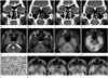
A 76-year-old man presented with a mass in the right nasal cavity that had developed 2 months ago. He had experienced right nasal stuffiness, hyposmia, and intermittent posterior nasal drip for 5 months. Approximately 2 months ago, a mass had been detected in the right nasal cavity by an otolaryngologist during rhinologic evaluation for the patient's nasal symptoms at a local medical clinic. The patient denied visual symptoms, and the motion of extraocular muscles was intact. Detailed ophthalmologic investigations were omitted by the otolaryngologist. His past history was unremarkable. Intranasal examination revealed a black, pigmented mass in the right nasal cavity, just below the right inferior turbinate. CT of the sinonasal area, performed using a Sensation 64 scanner (Siemens Healthcare, Forchheim, Germany) with a slice thickness of 2.0 mm revealed a fairly well-demarcated, homogeneously hyperattenuating (compared with brain) soft tissue mass involving the right lacrimal sac, nasolacrimal duct, and inferior meatus (Fig. 1A, B). The involved nasolacrimal duct was widened and eroded (Fig. 1A), and the inferior turbinate was elevated (Fig. 1B) by the mass. While the mass was in direct contact with orbital fat and with the medial wall of the right maxillary sinus and the lateral wall of the right anterior ethmoid sinus, there was no detectable invasion of other orbital structures, maxillary or anterior ethmoid sinus (Fig. 1A-D). The mass showed homogeneous, solid enhancement, following intravenous administration of contrast material (Fig. 1C, D). In view of CT features, our tentative diagnoses were lymphoma; primary mucosal melanoma; minor salivary gland tumor, including mucoepidermoid carcinoma and adenoid cystic carcinoma; and papillomas. Intranasal biopsy demonstrated mucosal melanoma. Preoperative MRI of the head and neck was performed by using a 3.0-T unit (Signa Excite; GE Medical System, Milwaukee, WI, USA). The mass was hypointense to gray matter on fast spin echo T2-weighted images (T2WI) [repetition time (TR)/echo time (TE) = 4000/106; echo train length = 22; matrix number/number of excitation = 512 × 224/2.0; slice thickness = 6.0 mm](Fig. 1E), and hyperintense to gray matter on spine echo (SE) T1-weighted images (T1WI) (TR/TE = 616.7/11.0; matrix number/number of excitation = 320 × 160/1.0; slice thickness = 6.0 mm) (Fig. 1F). Intense enhancement was noted on gadolinium-enhanced SE T1WI (TR/TE = 816.7/18.0; matrix number/number of excitation = 320 × 128/0.8; slice thickness = 8.0 mm) with fat saturation (Fig. 1G). Initial staging 18F-FDG PET/CT, performed by using Discovery STE (GE Healthcare, Milwaukee, WI, USA), demonstrated slightly increased fluorodeoxyglucose (FDG) uptake (maximum standardized uptake value = 3.2 g/mL) of the mass at the lacrimal sac (Fig. 1H) and inferior meatus. There were no findings of primary or metastatic melanoma elsewhere in the body on MRI and 18F-FDG PET/CT. He underwent en bloc resection of the mass in the lacrimal drainage apparatus via medial maxillectomy, partial ethmoidectomy, dacryocystectomy, and partial resection of medial orbital floor, lateral nasal wall, and nasal bone. Surgical margins were free of malignant cells. Histological examination revealed that the tumor was composed of large polygonal cells with prominent nucleoli and brownish melanin pigments (Fig. 1I). Immunohistochemically, tumor cells were positive for S-100 protein and HMB-45, consistent with malignant melanoma. The head and neck surgeon decided to follow-up without additional adjuvant radiotherapy, because he was confident of complete removal of the tumor and the surgical margins were negative for residual tumor. There were no definite findings of recurrence until 18 months after surgery at follow-up 18F-FDG PET/CT revealed faint FDG uptake at the anterior wall of the right maxillary sinus (Fig. 1J). He was given fractionated radiotherapy at a total dose of 40 Gy (500 cGy/fraction, 2 fractions/week for 4 weeks) directed at the anterior wall of his right maxillary sinus. Despite adjuvant radiotherapy, the recurrent disease progressed thereafter on periodic follow-up 18F-FDG PET/CT (Fig. 1J). The patient refused further management at our hospital and was lost to follow-up.
DISCUSSION
Our case represents primary mucosal melanoma of the lacrimal sac with extension into the inferior meatus through the nasolacrimal duct.
Primary mucosal melanoma, a very rare neoplasm accounting for 0.8-1.3% of all melanoma, most commonly occurs in the head and neck, mainly in the sinonasal area (5). Primary mucosal melanomas of the lacrimal drainage apparatus are extremely rare, and the majority of them arise from the lacrimal sac (1-4). Given that melanocytes are not usually present in the lacrimal sac, it has been hypothesized that primary mucosal melanoma of the lacrimal sac originates from the melanotic cells that may be inadvertently laid down beneath the lacrimal sac mucosa during embryogenesis or that may migrate into the lacrimal sac from adjacent conjunctiva (6, 7).
Early diagnosis of primary mucosal melanoma of the lacrimal drainage apparatus would yield the greatest chance of survival. However, it is not easy to distinguish primary mucosal melanoma of the lacrimal drainage apparatus from more common chronic dacryocystitis clinically. As such, imaging is important not only for the diagnosis but for the evaluation of the extent of primary mucosal melanoma of the lacrimal drainage apparatus. The Pujari et al. (1) reported a case of primary mucosal melanoma of the lacrimal sac, which was seen as a fairly well-defined, isoattenuating mass with invasion of the adjacent bone on CT. Sitole et al. (2) described CT features of a case of lacrimal sac melanoma, which was seen as a nonenhancing soft tissue mass of the left lacrimal sac with widening and erosion of the nasolacrimal duct. In contrast, Nam et al. (3) noticed that intense enhancement of the primary mucosal melanoma of the lacrimal sac on CT. In our case, solidly enhancing primary mucosal melanoma of the lacrimal drainage apparatus and bony changes of the nasolacrimal duct and inferior turbinate were well demonstrated on CT.
The differential diagnoses of a solidly enhancing mass involving the lacrimal drainage apparatus on CT include epithelial tumors comprising squamous cell carcinoma, transitional cell carcinoma, and minor salivary gland tumors (mucoepidermoid carcinoma and adenoid cystic carcinoma), and nonepithelial tumors comprising mesenchymal tumors, lymphoma (diffuse large B-cell lymphoma and extranodal marginal zone B-cell lymphoma of mucosa-associated lymphoid tissue), and primary mucosal melanoma. The literature reports 300-500 cases of tumors involving the lacrimal drainage apparatus, most as isolated case studies, with rare series (8). Because of the rarity of the tumors involving the lacrimal drainage apparatus, the characteristic imaging features of individual tumors of the lacrimal drainage apparatus have not been fully discussed. Our literature review reveals that the bony destruction and invasion of adjacent structures tend to be frequent in the squamous and transitional cell carcinomas, the two most common tumors involving the lacrimal drainage apparatus. In our case, the primary mucosal melanoma involving the lacrimal drainage apparatus was seen as a well-demarcated, solidly enhancing mass with bony remodeling, but without bony destruction or invasion of adjacent structures. The mass was homogeneously hyperattenuating, compared with brain parenchyma, and it showed homogeneous enhancement on contrast-enhanced CT. Accordingly, lymphoma, especially B-cell phenotype, and primary mucosal melanoma were the first diagnostic considerations. However, minor salivary gland tumors and papillomas could not be definitively excluded. We recommended MRI for the next step of evaluation.
Melanotic melanoma of the head and neck is expected to show high signal intensity (SI) on T1WI and low SI on T2WI owing to the paramagnetic property of melanin pigment, although hemorrhage and other substances may masquerade melanin, and amelanotic melanoma may not show such characteristic findings (9). In the one and only case report presenting MRI findings of malignant melanoma of the lacrimal sac, Billing et al. (4) noticed that the mass involving the lacrimal sac and nasolacrimal duct showed intermediate SI on both T1WI and T2WI, and was enhanced following intravenous administration of gadolinium. In our case of primary mucosal melanoma of the lacrimal drainage apparatus, MRI showed characteristic features of melanotic melanoma, in that the mass was hyperintense on T1WI and hypointense on T2WI, compared with gray matter, and intensely enhanced on Gd-enhanced T1WI, which thus helped us to predict a histological diagnosis.
18F-FDG PET/CT findings of primary mucosal melanoma of the lacrimal drainage apparatus have not been presented in the literature. Initial and periodic follow-up 18F-FDG PET/CT would enable initial staging and early detection of the locoregional recurrence and metastasis of the primary mucosal melanoma of the lacrimal drainage apparatus, as in other areas of the head and neck (10). In our case, the initial 18F-FDG PET/CT revealed a mass with mild FDG uptake. Recurrent disease occurring at the anterior wall of the right maxillary sinus was seen as a mass with mild FDG uptake on follow-up 18F-FDG PET/CT obtained 18 months after surgery, and progressive enlargement and increment of FDG uptake of the recurrent disease thereafter were excellently demonstrated on periodic follow-up 18F-FDG PET/CT.
In summary, imaging is crucial for early diagnosis of primary mucosal melanoma of the lacrimal drainage apparatus, and thus it improves the chance of patient survival. CT seems to be valuable for early detection of the mass and associated bony changes of the lacrimal drainage apparatus; MRI, for lesion characterization; and initial and follow-up 18F-FDG PET/CT, for initial staging and surveillance of recurrent and metastatic diseases following surgery, respectively.
 XML Download