

 PDF
PDF ePub
ePub Citation
 Print
Print


INTRODUCTION
Malignancy of the biliary hilum (e.g., hilar cholangiocarcinoma, and gallbladder cancer with hilar invasion) has an extremely poor prognosis, with a 5-year survival rate of less than 10% (1, 2). Most of the cases are known to be inoperable and the reported resectability rate of hilar bile duct tumor is approximately 10-20% (3). Thus, the majority of patients receive just palliative treatment.
Following the development of the T-configured dual stent, single percutaneous transhepatic approach for bilateral drainage has been widely used in the field of interventional radiology (4).
More recently, the endoscopic Y-stent, a novel variant of the T-configured stent, is being widely used for bilateral internal drainage of malignant hilar obstruction (5-8). The technical success rate and the patency of the endoscopic Y-stent are reported to be similar to those of percutaneous approaches (5).
Cotton et al. (9) defined an early complication as any stent-related complication which develops within 30 days of stent placement (9). Potential complications of endoscopic retrograde cholangiopancreatoscopy include biliary sepsis, hemorrhage, duodenal perforation, pancreatitis and early stent migration. Early obstruction with biliary sepsis can be particularly fatal.
Hwang et al. (6) reported that the early complication rate of endoscopic Y-stenting is 10%. In their study, all of the early complications observed were infectious ones and were accompanied by obstructive jaundice. And percutaneous transhepatic biliary drainage (PTBD) is required in cases of an infectious complication caused by obstructive jaundice.
In general, patients with stent malfunction undergo PTBD, and thus, internal drainage cannot be achieved.
To manage the early failure of endoscopic biliary stenting, we applied percutaneous transhepatic stenting as a rescue for internal drainage. The purpose of this study is to evaluate the feasibility and effectiveness of percutaneous transhepatic stenting as a rescue for the early occlusion of endoscopic biliary stents.
MATERIALS AND METHODS
We retrospectively studied ten patients (4 men and 6 women; age range, 52-78 years; mean age, 69 years) with inoperable biliary obstruction and early endoscopic Y-stent failure between August 2005 to November 2010. Early endoscopic stent failure was defined as any technical failure of endoscopic Y-stenting, with early complication development. We used the definition of early complications as suggested by Cotton et al. (9). They defined early complications as any stent-related complications that develop within 30 days. All of the patients included in this study had jaundice and fever caused by the endoscopic stent malfunction.
All patients were considered to be inappropriate for surgical resection on the basis of the tumor extent and their medical condition, and thus received endoscopic Y-stenting. Causes of early endoscopic stent failure included second stent (endoscopic Y-stent) insertion failure (n = 5) (Fig. 1), malposition of the second stent in B4 (n = 2) (Fig. 2), partial drainage due to an anatomical variation (n = 1) and tumor invagination through the stent mesh (n = 2). Mean time to endoscopic stent failure was 13.5 days (range 4-45 days). After failure of endoscopic stenting, all patients underwent PTBD procedures with the use of sonographic and fluoroscopic guidance. Rescue stents were inserted through the PTBD tract several days after the PTBD. The mean time of rescue stenting after PTBD was 7.3 days (range 2-13 days). Metallic stents were placed through the stenotic or obstruction lesions in all patients. The technique used to place stents is illustrated in Figs. 1 and 2.
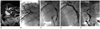
Endoscopic Y-stenting was performed in patient 1 who had malignant hilar obstruction due to gallbladder cancer with hilar invasion (Fig. 1A). But after the procedure, patient 1 had fever and jaundice caused by a second stent insertion failure. And on cholangiogram, obstruction at the right hepatic duct was identified (Fig. 1B).
0.035-inch 150-cm long hydrophilic guide wires (Terumo, Tokyo, Japan) were negotiated through the endoscopically induced strictures and stent meshes into the common bile duct (CBD), and both wires were dilated by a balloon catheter over the guide wire (Fig. 1C, D). Rescue stents were deployed through the mesh of the endoscopic stents and CBD. Following stent deployment, an 8.5-Fr drainage catheter (Uresil, Skokie, IL, USA) was placed over the guide wire. Three days after stent placement, contrast material was injected to confirm the position and patency of the rescue stents, and the PTBD catheters were removed (Fig. 1E).
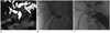
Obstructive jaundice after endoscopic Y-stenting occurred due to a second stent malposition in patient 2. And rescue stenting at the right hepatic duct was performed with the same technique described as above (Fig. 2).
Broad spectrum antibiotics were given to patients before and after each procedure. All of the rescue stenting was performd by one experienced interventional radiologist (C.W.K.).
The outcome of rescue stenting was evaluated using the following parameters: technical success (successful stent insertion), clinical success (successful drainage), procedure-related mortality, 30-day mortality, patency time and survival time.
We applied the criteria which Hwang et al. (6) used in their evaluation of the procedure. Technical success was defined by both successful stent placement and resolution of the biliary stenosis or occlusion, with copious flow of the contrast medium through the stent. Clinical success was defined as a significant fall in the bilirubin level to less than a third of the level before the procedure within the previous two weeks. Procedure-related mortality was defined as death directly related to complications of stent insertion. Patency time was defined as the number of days from the second drainage to reintervention or the patient's death. Survival time was defined as the interval between the time of rescue stenting and the patient's death.
Statistical analyses were performed using commercially available software (SPSS 10.0; SPSS Inc., Chicago, IL, USA). The rates of stent patency and survival were calculated using the Kaplan-Meier method.
Informed consent was obtained from all patients before the procedures. Certification of the Institutional Review Board was waived for this retrospective study.
RESULTS
In this study, two patients had gallbladder cancer with hilar invasion and eight patients had a Klatskin tumor. The cases were classified as either type II (n = 2), type IIIa (n = 4) or type IV (n = 4) according to the Bismuth classification.
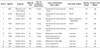
The results of all of the cases were consistent with the definition of early endoscopic stent failure. Mean time to endoscopic stent failure was 13.5 days (range 4-45 days). Causes of early endoscopic stent failure included second stent (endoscopic Y-stent) insertion failure (n = 5) (Fig. 1), malposition of the second stent in B4 (n = 2) (Fig. 2), partial drainage due to an anatomical variation (n = 1) and tumor invagination through the stent mesh (n = 2). Tumor invagination was defined as narrowing of the stent lumen on cholangiogram, and tumor within the stent lumen as seen on CT or MR. The mean time of rescue stenting after PTBD was 7.3 days (range 2-13 days) (Table 1).
Uncovered self-expandable metal stents were used in nine patients, and in one patient, a balloon expandable stent was used. Luminexx stent (Bard, Murray Hill, NJ, USA) (n = 5), Niti-D (Taewoong, Seoul, Korea) (n = 3), Hercules DH (S&G biotech, Seongnam, Korea) (n = 1), and Express LD balloon expandable stent (Boston Scientific Corp, Natick, MA, USA) (n = 1) were used.
The placement of the rescue stent was technically and clinically successful in all of the patients (technical and clinical success rate, 100%). On cholangiogram, copious flow of the contrast medium through the stent was observed in all of the patients who underwent rescue stenting. And within 2 weeks after rescue stenting, the serum bilirubin level dropped to less than one-third of the level before the procedure. There were no procedure-related deaths, and the 30-day mortality rate following stent insertion was 10% (patient 8, due to cardiac arrest). The mean patency time of rescue stenting was 122 days [range 20-330 days, 95% confidential interval (CI), 50.8-193.2 days]. And the median patency time was 80 days (95% CI, 28.3-131.6 days). The mean survival time from rescue stenting was 226.3 days (range 30-635 days, 95% CI, 85.7-366.9 days). And the median survival time was 121 days (95% CI, 74.5-167.5 days). The last patient (patient 10) is currently still alive. The time interval between the time of rescue stenting and the end of the study for the last patient (patient 10) was 191 days.
DISCUSSION
Since the development of the T-configured stent and endoscopic Y-stent, an increasing number of endoscopists have started using the Y-stent for malignant hilar obstruction. There are several reports supporting the usefulness of endoscopic management (5-8). However, early endoscopic stent malfunction and unresolved biliary obstruction potentially lead to life-threatening conditions.
To the best of our knowledge, this is the first report of a PTBD with internal drainage for the management of early endoscopic Y-stent failure in hilar tumor.
Standard techniques used to treat this problem include inserting endoscopic plastic stents and PTBD. But the patency of plastic stents lasts for a shorter time than that of metallic stents, and the insertion of a plastic stent through the metal stent mesh is very difficult. PTBD is a relatively simple method of redrainage in hilar obstruction but it has several disadvantages compared to internal drainage; the major disadvantages are loss of body fluid volume/electrolytes, pain, and inconvenience. They can act as risk factors for many fatal complications in terminal patients. And PTBD is far more inconvenient than other techniques for patients, and also an external catheter causes pain (10).
Ridtitid et al. (11) reported a second type of intervention, with an endoscopic self-expandable metallic stent, in patients with malignant biliary obstruction (including both hilar and non-hilar tumors). The study reported the performance and feasibility of the second endoscopic self-expandable metallic stent. And they compared the results of the second endoscopic metallic stents with those of PTBD and plastic stents. And second endoscopic metallic stents showed some effectiveness in non-hilar obstruction. But the results of second endoscopic metallic stent insertion in hilar obstructed patients (n = 3) showed short survival and patency (median patency 60 days, median survival 215 days) although it was technically successful (technical success rate, 100%).
Compared to the results of Ridtitid's reports (11) in hilar-obstructed patients, the results of our study showed a relatively longer patency (median patency 80 days). And the median survival time was shorter (median survival 121 days). The shorter survival time is believed to be due to the higher proportion of patients with high grade hilar obstruction in our study. Because we only included those patients with early stent failure, a simple comparison of the survival times was not appropriate.
The results of our rescue stenting showed shorter patency and survival times in comparison with the results of primary transhepatic stenting for malignant hilar obstruction using T-configured dual stents (mean patency 170.3 days) (4). There were several causes of the worse outcome in rescue stenting. In this study, the most common cause of early failure in endoscopic Y-stenting was technical problems, such as second stent insertion failure, stent malposition and partial drainage due to anatomical variation. It seems that the unsuitable positioning of the primary endoscopic stents due to technical problems led to the worse outcome. Rescue stenting itself is technically more difficult than primary transhepatic stenting. Also most of the patients with early endoscopic Y-stents already had infectious complications (such as infectious cholangitis) by the time they received the rescue stents, which could have also contributed to the differences in the outcome.
Transhepatic rescue stenting is a kind of new procedure. And it was technically feasible in our study. There is no other solution for early failure of endoscopic stents except for a PTBD procedure. Though rescue stenting has no definite survival benefit, it makes internal drainage possible after the early failure of endoscopic stenting. So transhepatic rescue stenting can be a good alternative choice with respect to improvement in the quality of life.
Endoscopic metal stent placement is now generally accepted as the palliative treatment of choice in patients with unresectable hilar cholangiocarcinoma. In Bismuth types I and II, endoscopic stenting is a safe and feasible method. However in Bismuth types III and IV, the results have not always been satisfactory. According to Paik et al. (12), the rate of successful biliary decompression in high grade obstruction such as Bismuth types III and IV was significantly higher in the percutaneous approach than in the endoscopic approach (12). All things taken together, primary transhepatic hilar stenting is a better choice for high grade malignant hilar obstruction.
This study has some limitations. First, the data were collected retrospectively. Second, the causes resulting in malignant hilar obstruction in this study were multiple (gall bladder cancer and Klastskin tumor). Third, a variety of stent products were used. Fourth, a small number of patients with early endoscopic stent failure were enrolled in this study, so a large group study is mandatory in the future.
In early failure of endoscopic biliary stenting, percutaneous transhepatic recanalization can be a possible solution. However, the stent patency time and patient's survival time were shorter than those of primary percutaneous stenting.
 XML Download