

 PDF
PDF ePub
ePub Citation
 Print
Print


INTRODUCTION
The os supratalare is quite a rare accessory bone located along the superior surface of the ridge along the talar head/neck, and may be seen distally over the head (1, 2). Only a few studies were published focusing on the incidences of this accessory ossicle. According to previous reports, the incidence of os supratalare depicted on radiography was ranged from 0.2% to 2.4% (2-4). The os supratalare, as most accessory ossicles of the ankle and foot, usually remain asymptomatic and are incidentally identified in radiographs obtained for other reasons (1). However, they can also cause pains or degenerative changes in response to overuse and trauma (5).
In this case report, we present the radiography, computed tomography (CT) and magnetic resonance imaging (MRI) manifestations of a symptomatic os supratalare in a 21-year-old woman. To the best of our knowledge, there have not been any previously reported imaging findings of symptomatic os supratalare.
CASE REPORT
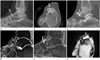
A 21-year-old woman was presented with a history of right dorsal hind foot pain associated with hard bump for one month duration. She had visited the local clinic and was referred to our hospital under the impression that the lesion of the dorsal hind foot was an osteogenic tumor. There was no history of trauma. Physical examinations revealed hard masses with tenderness in the area of dorsal surface of talar head. Lateral radiograph of the right ankle revealed a bony protuberance of the dorsal aspect on the right talus and a subtle radiolucent line in the bony protuberance along the dorsal aspect of the talar head (Fig. 1A). CT axial and sagittal images with 2.0 mm section demonstrated an accessory ossicle partially separated from dorsal aspect of the talar head. A narrow and mild irregular interface between accessory ossicle and the talar head with several small cysts and sclerotic changes along the interface, and small bony productive changes were also noted (Fig. 1B, C). Subsequently, MRI for the right ankle was performed with 3.0 T unit (Achieva, Philips Healthcare, Best, the Netherlands). A T2-weighted fast spin-echo image (4368.4/100.0) was obtained in the axial plane. A spin echo proton density (PD) spectral pre-saturation with inversion recovery (SPIR) sequence (2000.0/30.0) was obtained in the sagittal plane. T1-weighted SPIR images were also obtained in the sagittal plane (785.7/20.0) after IV administration of gadopentetate dimeglumine (Magnevist®, Bayer Schering Pharma AG, Berlin, Germany) 0.1 mmol/L per kilogram of body weight. The slice section thickness was 3 mm and the field view was 150 × 150 mm. Mild hyperintense signal intensities and enhancements in the accessory bone were noted on PD SPIR images and gadolinium-enhanced T1-weighted SPIR images, respectively (Fig. 1D, E). These findings were interpreted as a bone marrow edema. A peripheral enhancing multiloculated cystic lesion was incidentally revealed in the anterior aspect of the os supratalare (Fig. 1E, F). This cystic lesion was interpreted as a ganglion cyst or an adventitial bursa. On the basis of these imaging findings, the symptomatic os supratalare was diagnosed, due to degenerative changes across its synchondrosis together with cystic lesions in the anterior aspects of the os supratalare. The patient was treated with conservative managements including restricted physical activities with applications of short leg splint on an outpatient basis. But, the patient refused surgery and further follow-ups.
DISCUSSION
Most accessory ossicles of the ankle and foot usually remain asymptomatic, and are incidentally identified in obtained radiographs (1). However, they can cause pains or degenerative changes in response to overuse, trauma and irritation of the overlying soft tissues (5). Moreover, they can simulate other entities such as fractures on radiography (6). Many symptomatic accessory ossicles of the ankle and foot in various reports have already been discussed. However, imaging findings of painful os supratalare has not yet been reported.
Traditionally, the accessory ossicles have been evaluated by means of radiography or scintigraphy. Recently, additional CT and MRI have been considered as useful modalities to distinguish accessory ossicles from their mimickers such as fractures or bone tumors (5). In our case, lateral radiographs revealed a bony protuberance on the dorsal aspect of the talus. Only a subtle radiolucent gap between the os supratalare and talus was demonstrated on the radiography. This imaging finding might have led to misdiagnose of the os supratalare as osteogenic tumor by the referred physician. In our case, CT utilizing the conventional axial, coronal and sagittal planes clearly showed the location and relationship of the os supratalare. The os supratalare was partially fused with the talus at its medial part. Os supratalare, as other accessory ossicles, may be fused with talus or remain as a free accessory bone (1). CT could optimally show sclerosis and several degenerative small cystic lesions along the interface between the os supratalare and the talus. These may indicate chronic stresses with resultant injuries to the synchondrosis (7), in our case, the histological correlations could not be obtained because surgical excision was not performed. Therefore, the mild hyperintense signal intensity line between the os supratalare and the talar head was demonstrated on the fat suppression images. This finding may reflect the fibrocartilagenous nature of the synchondrosis (6) or the fluid within the disrupted synchondrosis (7). Bone marrow edema in os supratalare and mild irregularity along the interface between the os supratalare and talus were also demonstrated on MRI. Similar MRI findings had been reported in os sustentaculi and accessory navicular bone. According to their reports, these findings can be interpreted as degenerative/reparative changes due to chronic stresses at the synchondrosis and the source of pain (6, 8).
In this case, osteochondroma are excluded because of no perceptible cartilaginous cap on the MRI. The os supratalare should not be confused with old fractures of the talar beak, talar ridge or osteophyte. From the radiological standpoint, fracture represents more sharp and more irregular interfaces, and also the lack of complete cortication (5). The os supratalare presented in our report showed only mild irregular interface between talus and well-cortication. Nonetheless, imaging distinctions alone may be difficult. Thus, clinical history should be kept in mind (5). In our case, an old fracture of talar beak, talar ridge or osteophyte was considered as unlikely due to complete absence of prior trauma.
In our case, the multiloculated cystic lesion in the anterior aspect of the os supratalare was incidentally revealed on MRI. This cystic lesion was interpreted as a ganglion or an adventitial bursa. Osuji and McAdams (9) had reported a case of a ganglion cyst associated with os ulnostyloideum. In their case, the ganglion cyst originated from a pseudarthrosis between the ulnar styloid process and the os ulnostyloideum. Adventitial bursa can also be developed by accessory ossicles (10). It may be caused by repeated irritations of adjacent soft tissues by the accessory ossicles and is suggestive of chronic lesions rather than acute traumatic conditions. Although in our case, histological correlation could not be obtained and origin of the cystic lesion could not be confirmed, symptoms may be attributed to os supratalare together with the cystic lesion.
Herein, MRI is the useful imaging modality due to its ability of demonstrating bone marrow abnormalities and hyperintense signal intensities between the accessory ossicles and the talus. Moreover, cystic lesion in the anterior aspect of the os supratalare was also well-demonstrated because MRI provides excellent soft-tissues contrast.
In conclusion, an os supratalare is a rare reason for dorsal foot pains that may be depicted on CT or MRI. The CT and MRI are helpful to distinguish it from a fracture or an osteochondroma. Nevertheless, MRI can provide additional information on associated soft tissue lesions. Increased knowledge on imaging findings and clinical significances of os supratalare will be helpful for accurate diagnosis and appropriate management.
 XML Download