

 PDF
PDF ePub
ePub Citation
 Print
Print


Abstract
Purpose
Thyroid pyramidal lobe (TPL) is a normal variant of the thyroid gland, but few imaging studies of TPL have been published. The purpose of this study is to investigate the frequency, location, size (length, maximal AP diameter, maximal transverse diameter), and upper end level of TPL with its separation from the main thyroid gland on preoperative neck CT and to compare them with operative findings in order to assess the diagnostic accuracy of neck CT for detection TPL.
Materials and Methods
46 patients, who underwent preoperative neck CT before thyroidectomy, were included in the study. The frequency, location, size, and upper end level of TPL with its separation from the main thyroid gland on the neck CT was analyzed by a single radiologist.
Results
The sensitivity, specificity, positive predictive value, negative predictive value and accuracy of neck CT for detecting TPL was 77.8%, 89.5%, 91.3%, 73.9% and 82.6%. There was a significant difference in maximal AP diameter, location, upper end level, and its separation from main thyroid gland between CT and operative findings (p < 0.05), but there was no significant difference in the length and maximal transverse diameter of TPL (p > 0.05).
Figures and Tables
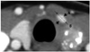 | Fig. 1Size measurement of thyroid pyramidal lobe in CT image on a picture archiving and communication system. Maximal anteroposterior (A) and transverse (B) diameters of thyroid pyramidal lobe are measured in axial images. The length of thyroid pyramidal lobe are determined according to the number of axial images showing thyroid pyramidal lobe.
|
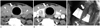 | Fig. 2A 52-year-old man with a right pyramidal lobe detected on neck CT and in surgery. Nonenhanced axial (A) and contrast-enhanced axial (B) CT images show the right pyramidal lobe (arrow) with the same attenuation and enhancement as the main thyroid gland (arrowheads). The right pyramidal lobe (C) is observed during thyroid surgery (arrow).
|
References
1. Moon WJ, Baek JH, Jung SL, Kim DW, Kim EK, Kim JY, et al. Ultrasonography and the ultrasound-based management of thyroid nodules: consensus statement and recommendations. Korean J Radiol. 2011; 12:1–14.
2. Hay ID, Thompson GB, Grant CS, Bergstralh EJ, Dvorak CE, Gorman CA, et al. Papillary thyroid carcinoma managed at the Mayo Clinic during six decades (1940-1999): temporal trends in initial therapy and long-term outcome in 2444 consecutively treated patients. World J Surg. 2002; 26:879–885.
3. Mazzaferri EL, Kloos RT. Clinical review 128: Current approaches to primary therapy for papillary and follicular thyroid cancer. J Clin Endocrinol Metab. 2001; 86:1447–1463.
4. Pacini F. Follow-up of differentiated thyroid cancer. Eur J Nucl Med Mol Imaging. 2002; 29:Suppl 2. S492–S496.
5. Hollinshead WH. The head and the neck. Anatomy for surgeons. NewYork: Hoeber-Harper;1961. vol 2:p. 517–531.
6. Ranade AV, Rai R, Pai MM, Nayak SR, Prakash , Krisnamurthy A, et al. Anatomical variations of the thyroid gland: possible surgical implications. Singapore Med J. 2008; 49:831–834.
7. Joshi SD, Joshi SS, Daimi SR, Athavale SA. The thyroid gland and its variations: a cadaveric study. Folia Morphol (Warsz). 2010; 69:47–50.
8. Park JY, Kim DW, Park JS, Kang T, Kim YW. The prevalence and features of thyroid pyramidal lobes as assessed by computed tomography. Thyroid. 2012; 22:173–177.
9. Kim DW, Jung SL, Baek JH, Kim J, Ryu JH, Na DG, et al. The prevalence and features of thyroid pyramidal lobe, accessory thyroid, and ectopic thyroid as assessed by computed tomography: a multicenter study. Thyroid. 2013; 23:84–91.
10. Andersen PE, Kinsella J, Loree TR, Shaha AR, Shah JP. Differentiated carcinoma of the thyroid with extrathyroidal extension. Am J Surg. 1995; 170:467–470.
11. Tsang RW, Brierley JD, Simpson WJ, Panzarella T, Gospodarowicz MK, Sutcliffe SB. The effects of surgery, radioiodine, and external radiation therapy on the clinical outcome of patients with differentiated thyroid carcinoma. Cancer. 1998; 82:375–388.
12. Pacini F, Schlumberger M, Harmer C, Berg GG, Cohen O, Duntas L, et al. Post-surgical use of radioiodine (131I) in patients with papillary and follicular thyroid cancer and the issue of remnant ablation: a consensus report. Eur J Endocrinol. 2005; 153:651–659.
13. Moore KL, Persaud TVN. The developing human: clinically oriented embryology. Philadelphia: WB Saunders Company;1993. p. 200–203.
14. Won HS, Chung IH. Morphologic variations of the thyroid gland in Korean adults. Korean J Phys Anthropol. 2002; 15:119–125.
 XML Download