

 PDF
PDF ePub
ePub Citation
 Print
Print


Abstract
Purpose
To investigate a new image acquisition method [four dimensional T1-weighted high resolution imaging with volume excitation (4D THRIVE)] which enables an accurate hepatic arterial phase definition. The feasibility and its potential for detection and characterizing focal liver lesions (FLLs) are being evaluated.
Materials and Methods
115 FLLs underwent liver MRI that included the 4D THRIVE-contrast enhanced timing robust acquisition order (CENTRA)-keyhole sequence. Triple arterial phase was obtained during a single breath-hold. Images were reviewed for image quality, lesion conspicuity, and lesion detection. Two radiologists independently assessed images from phase I, II, III and through the triple arterial phase, which were all reviewed separately and in random order. The image quality was scored by using the five-point scale, and then, one phase for lesion with greatest conspicuity was selected. The enhancement pattern for FLLs was analyzed.
Results
The detection rate was the highest on phase III. The image quality was greater than grade 3 with fair inter-observer agreements. The phase III showed greater conspicuity than phase I and II. Hepatocellular carcinomas (n = 38) showed variable enhancement pattern. Metastasis (n = 14) showed rim enhancement (n = 6), homogenous (n = 3) and no enhancement (n = 5). Most hemangiomas demonstrated homogenous enhancement (6/9, 67%).
Figures and Tables
Fig. 1
Schematic image of the keyhole technique (A) and the alternating viewsharing technique (B). The peripheral k-space data, called reference, is acquired only in last arterial phase and is shared with every phase. The central k-space data is determined by the keyhole percentage. In four dimensional T1-weighted high resolution imaging with volume excitation, the central ky-kz disc (keyhole) is subdivided in three regions, P+ (positive peripheral region), C (central region) and P- (negative peripheral region). The central region is acquired every arterial phase, but P+ and P- are shared with subsequent phase according to alternating viewsharing scheme as shown above. The view sharing percentage is determined by the rate of the area occupied by P+ (or P-) and C to the whole central disc.
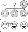
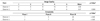
Fig. 2
A timing diagram shows the sequence of T1 weighted image acquisition. First, T1 weighted image without contrast injection is acquired within one breath hold. With short time of interval, contrast injection starts via peripheral venous route after delivering a message. 2D fluoroscopic real time imaging which is followed by contrast administration shows right atrium as we selected. When the contrast reaches right atrium, the MRI technologist manually presses the button to cessation of real-time display and to initiate breath-holding instructions. With one breath hold, three arterial phases are acquired with 4D THRIVE technique. According to alternating view sharing scheme as described, central disc of three phases are acquired first, and finally peripheral k-space date is acquired.
Note.-4D THRIVE = four dimensional T1-weighted high resolution imaging with volume excitation
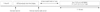
Fig. 3
Small HCCs in two different patients. From left to right, dynamic T1 weighted MRI images from three consecutive arterial phases, portal phase and delayed phase are shown.
A. Images from 49 years old male patient demonstrate a small hepatic nodule with gradual homogeneous arterial enhancement and delayed washout (white arrows).
B. In images from another 59 years old male patient with HCC show heterogeneous enhancement (black arrows).
Note.-Delay = 20 minutes delayed phase, HCC = hepatocellular carcinoma, PP = portal phase
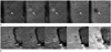
Fig. 4
Surgically confirmed hepatic metastasis from colon cancer in 57 years old patient. In arterial phase, the lesion shows peripheral rim enhancement. There is no definite difference of degree of contrast enhancement between each arterial phase.
Note.-Delay = 20 minutes delayed phase, PP = portal phase
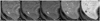
Fig. 5
Small hemangiomas in two different patients.
A. Dynamic T1 weighted contrast enhanced dynamic MRI shows typical peripheral nodular and centripetal enhancement of hemangioma.
B. Dynamic MRI images from another patient with small hemangioma show homogeneous enhancement. Note the degree of enhancement is same as that of adjacent hepatic arteries.
Note.-Delay = 20 minutes delayed phase, PP = portal phase
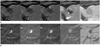
Fig. 6
56 years old male patient with increased peripheral eosinophilic count. Portal phase CT (A) shows ill defined low density lesion (arrow). After 4 months, the lesion was disappeared on follow-up CT (B) and eosinophilic count was normalized. Dynamic MRI (C) shows subtle homogeneous enhancing lesion on arterial phase. And this lesion shows low signal intensity when compared with adjacent liver parenchyma on delayed phase (arrows).
Note.-Delay = 20 minutes delayed phase, PP = portal phase
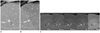
Fig. 7
Marked ringing artifact induced by respiratory motion. The artifact is shown on equally both phase I (A) and phase III (B), obscuring underlying focal liver lesion. A low signal intensity focal liver lesion is shown in delayed phase image (C) which is acquired during another breath hold.
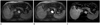
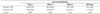
Table 1
Standards of Reference for Focal Liver Lesions
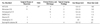
Note.-*27 of 29 lesions were diagnosed by AASLD guideline. The two remaining lesions were not satisfied with AASLD guideline initially, but showed lipiodol uptake on TACE.
†2 lesions were increased in size on follow-up image, and the other lesions were diagnosed by concurrent serum AFP increase (> 200 ng/mL).
AASLD = American association for the study of liver diseases, AFP = alpha fetoprotein, DN = dysplastic nodule, F/U = follow up, FLL = focal liver lesion, HCC = hepatocellular carcinoma, RN = regenerative nodule, TACE = transarterial chemoembolization
References
1. Hawighorst H, Schoenberg SO, Knopp MV, Essig M, Miltner P, van Kaick G. Hepatic lesions: morphologic and functional characterization with multiphase breath-hold 3D gadolinium-enhanced MR angiography--initial results. Radiology. 1999; 210:89–96.
2. Elsayes KM, Narra VR, Yin Y, Mukundan G, Lammle M, Brown JJ. Focal hepatic lesions: diagnostic value of enhancement pattern approach with contrast-enhanced 3D gradient-echo MR imaging. Radiographics. 2005; 25:1299–1320.
3. Kanematsu M, Semelka RC, Matsuo M, Kondo H, Enya M, Goshima S, et al. Gadolinium-enhanced MR imaging of the liver: optimizing imaging delay for hepatic arterial and portal venous phases--a prospective randomized study in patients with chronic liver damage. Radiology. 2002; 225:407–415.
4. Goshima S, Kanematsu M, Kondo H, Yokoyama R, Miyoshi T, Nishibori H, et al. MDCT of the liver and hypervascular hepatocellular carcinomas: optimizing scan delays for bolus-tracking techniques of hepatic arterial and portal venous phases. AJR Am J Roentgenol. 2006; 187:W25–W32.
5. Hong HS, Kim HS, Kim MJ, De Becker J, Mitchell DG, Kanematsu M. Single breath-hold multiarterial dynamic MRI of the liver at 3T using a 3D fat-suppressed keyhole technique. J Magn Reson Imaging. 2008; 28:396–402.
6. Low RN, Bayram E, Panchal NJ, Estkowski L. High-resolution double arterial phase hepatic MRI using adaptive 2D centric view ordering: initial clinical experience. AJR Am J Roentgenol. 2010; 194:947–956.
7. Lee VS, Lavelle MT, Rofsky NM, Laub G, Thomasson DM, Krinsky GA, et al. Hepatic MR imaging with a dynamic contrast-enhanced isotropic volumetric interpolated breath-hold examination: feasibility, reproducibility, and technical quality. Radiology. 2000; 215:365–372.
8. Beck GM, De Becker J, Jones AC, von Falkenhausen M, Willinek WA, Gieseke J. Contrast-enhanced timing robust acquisition order with a preparation of the longitudinal signal component (CENTRA plus) for 3D contrast-enhanced abdominal imaging. J Magn Reson Imaging. 2008; 27:1461–1467.
9. Bruix J, Sherman M. Practice Guidelines Committee. American Association for the Study of Liver Diseases. Management of hepatocellular carcinoma. Hepatology. 2005; 42:1208–1236.
10. Coenegrachts K, Ghekiere J, Denolin V, Gabriele B, Hérigault G, Haspeslagh M, et al. Perfusion maps of the whole liver based on high temporal and spatial resolution contrast-enhanced MRI (4D THRIVE): feasibility and initial results in focal liver lesions. Eur J Radiol. 2010; 74:529–535.
11. Cantwell CP, Setty BN, Holalkere N, Sahani DV, Fischman AJ, Blake MA. Liver lesion detection and characterization in patients with colorectal cancer: a comparison of low radiation dose non-enhanced PET/CT, contrast-enhanced PET/CT, and liver MRI. J Comput Assist Tomogr. 2008; 32:738–744.
12. Parikh T, Drew SJ, Lee VS, Wong S, Hecht EM, Babb JS, et al. Focal liver lesion detection and characterization with diffusion-weighted MR imaging: comparison with standard breath-hold T2-weighted imaging. Radiology. 2008; 246:812–822.
13. Kim KW, Kim AY, Kim TK, Park SH, Kim HJ, Lee YK, et al. Small (<or= 2 cm) hepatic lesions in colorectal cancer patients: detection and characterization on mangafodipir trisodium-enhanced MRI. AJR Am J Roentgenol. 2004; 182:1233–1240.
14. Davenport MS, Viglianti BL, Al-Hawary MM, Caoili EM, Kaza RK, Liu PS, et al. Comparison of acute transient dyspnea after intravenous administration of gadoxetate disodium and gadobenate dimeglumine: effect on arterial phase image quality. Radiology. 2013; 266:452–461.
15. Tanimoto A, Higuchi N, Ueno A. Reduction of ringing artifacts in the arterial phase of gadoxetic acid-enhanced dynamic MR imaging. Magn Reson Med Sci. 2012; 11:91–97.
16. Frederick MG, McElaney BL, Singer A, Park KS, Paulson EK, McGee SG, et al. Timing of parenchymal enhancement on dual-phase dynamic helical CT of the liver: how long does the hepatic arterial phase predominate? AJR Am J Roentgenol. 1996; 166:1305–1310.
17. Sharma P, Kalb B, Kitajima HD, Salman KN, Burrow B, Ray GL, et al. Optimization of single injection liver arterial phase gadolinium enhanced MRI using bolus track real-time imaging. J Magn Reson Imaging. 2011; 33:110–118.
18. Yoshioka H, Takahashi N, Yamaguchi M, Lou D, Saida Y, Itai Y. Double arterial phase dynamic MRI with sensitivity encoding (SENSE) for hypervascular hepatocellular carcinomas. J Magn Reson Imaging. 2002; 16:259–266.
19. Ito K, Fujita T, Shimizu A, Koike S, Sasaki K, Matsunaga N, et al. Multiarterial phase dynamic MRI of small early enhancing hepatic lesions in cirrhosis or chronic hepatitis: differentiating between hypervascular hepatocellular carcinomas and pseudolesions. AJR Am J Roentgenol. 2004; 183:699–705.
20. Nino-Murcia M, Olcott EW, Jeffrey RB Jr, Lamm RL, Beaulieu CF, Jain KA. Focal liver lesions: pattern-based classification scheme for enhancement at arterial phase CT. Radiology. 2000; 215:746–751.
21. Danet IM, Semelka RC, Leonardou P, Braga L, Vaidean G, Woosley JT, et al. Spectrum of MRI appearances of untreated metastases of the liver. AJR Am J Roentgenol. 2003; 181:809–817.
22. Yu JS, Rofsky NM. Hepatic metastases: perilesional enhancement on dynamic MRI. AJR Am J Roentgenol. 2006; 186:1051–1058.
23. Vilgrain V, Boulos L, Vullierme MP, Denys A, Terris B, Menu Y. Imaging of atypical hemangiomas of the liver with pathologic correlation. Radiographics. 2000; 20:379–397.
24. Byun JH, Yang DH, Yoon SE, Won HJ, Shin YM, Jeong YY, et al. Contrast-enhancing hepatic eosinophilic abscess during the hepatic arterial phase: a mimic of hepatocellular carcinoma. AJR Am J Roentgenol. 2006; 186:168–173.
 XML Download