

 PDF
PDF ePub
ePub Citation
 Print
Print


INTRODUCTION
Neuroendocrine neoplasms account for 1.2-1.5% of all gastrointestinal neoplasms, with an annual incidence of 1.6-2.0 new cases for every 100000 persons. These neoplasms may involve many different organs and sites, with a majority of the tumors occuring in small bowel, appendix, rectum, and the stomach (1). Biliary neuroendocrine neoplasms are extremely rare, and represents only 1% of all gastrointestinal neuroendocrine neoplasms. This is probably due to a very low numbers of neuroendocrine (Kulchitsky) cells in the biliary epithelium (2, 3). Because of its extremely rare incidence and presenting symptoms like painless jaundice, most extrahepatic biliary neuroendocrine neoplasms are misdiagnosed as adenocarcinoma prior to surgery (4). Most studies on these tumors consist of case reports with literature review and focus on histologic features or clinical outcomes.
This case is unique in that the role of imaging study in the diagnosis of neuroendocrine neoplasm of biliary duct has not been reported until now.
CASE REPORT
A 64-year-old man with a known diagnosis of pneumoconiosis was transferred to our hospital due to indigestion and jaundice that persisted for 3 weeks. The initial laboratory studies detected abnormally elevated serum levels of the total bilirubin and direct bilirubin (11.67 mg/dL and 8.37 mg/dL, respectively) and an elevated alkaline phosphatase level of 657 IU/L. Tumor markers, such as carcinoembryonic antigen, and CA19-9, were found to be within the normal range.
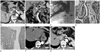
A contrast-media enhanced abdominal CT (Fig. 1A, B) detected a well-enhancing 2.0 cm polypoid mass in the proximal common bile duct (CBD), which had resulted in extrahepatic biliary obstruction. The mass demonstrated about 100 Hounsfield unit (HU) net enhancement (17 HU on pre-enhancement image, 113 HU on post-enhancement image) during the late arterial phase. Several enlarged lymph nodes were seen around the common bile duct, porta hepatis, and paraaortic space. Seven days after the CT scan, the patient underwent percutaneous transhepatic biliary drainage (PTBD) procedure. The percutaneous tubography revealed a small expansile intraductal polypoid mass in the proximal CBD with moderate dilatation of the intrahepatic ducts (Fig. 1C). Positron emission tomography (PET) CT showed a faint fluorodeoxyglucose (FDG) uptake [standardized uptake value (SUV) max: 2.4] in this mass.
Seven days subsequent to PTBD, CBD segmental resection and choledochojejunostomy was performed under a presumed diagnosis of cholangiocarcinoma. Gross examination revealed a 12.0 × 2.0 cm polypoid mass. Microscopically, the tumor had invaded beyond the wall of CBD to the surrounding adipose tissue with one regional lymph node metastasis. Histological examination (Fig. 1D, E) revealed the tumor to have both adenocarcinomatous component and neuroendocrine tumor. Immunohistochemistry was positive for COX-2, suggestive of adenomatous component, and also revealed positive expression of CD56, chromogranin, neuron-specific enolase, suggesting neuroendocrine component. The Ki67 labelling index, a proliferative marker, was 95%. Thus, the final diagnosis was confirmed as mixed endocrine-exocrine carcinoma.
The postoperative course was uneventful, until a follow-up abdominal CT (Fig. 1F, G) at 7 months revealed multiple nodules in both hepatic lobes. These lesions were small, arterial enhancing, and demonstrated subtle delayed washout. This imaging study was suggestive of hepatocellular carcinomas. On ultrasonographic exam, these masses were homogenous hyperechoic nodules in the liver parenchyma. Sono-guided liver biopsy was performed for several lesions. The pathology was confirmed to be metastastic mixed endocrine-exocrine carcinoma. The patient is currently alive undergoing chemotherapy treatment.
DISCUSSION
On one hand, biliary neuroendocrine neoplasms are extremely rare and represent only 1% of all gastrointestinal neuroendocrine neoplasms. On the other hand, adenocarcinoma is the most common type of all biliary duct tumors, accounting for approximately 80% of occurrence (2, 5). The most common presenting symptom of biliary neuroendocrine neoplasms is jaundice, and the most common sites are the common bile duct (58%), perihilar region (28%), cystic duct (11%), and the common hepatic duct (3%) (3). Most biliary neuroendocrine neoplasms are misdiagnosed before as adenocarcinoma preoperatively because of its extremely rare incidence and its similarity to the presenting symptoms and nonspecific radiologic findings of adenocarcinoma (4). The presenting symptoms involve jaundice, biliary colic pain, abdominal discomfort, and weight loss. Unlike those found in other locations, neuroendocrine neoplasms of the extrahepatic biliary ducts tend to have an indolent biological behavior, even when metastatic. This makes it more difficult for a correct diagnosis. The variety of nomenclature used for neuroendocrine neoplasm of the gastrointestinal tract add to the confusion among clinicians and radiologists.
Recently, a new classification of neuroendocrine neoplasm has been proposed and adopted by the World Health Organization (WHO). The WHO 2010 classification identified following histopathologic subtypes: 1) neuroendocrine tumor (NET) G1 and G2, which is defined as a well differentiated neuroendocrine neoplasm; 2) neuroendocrine carcinoma (NEC), which is a poorly differentiated, high grade malignant neoplasm; and, 3) mixed adenoneuroendocrine (exocrine endocrine) carcinoma, which has a morphologic phenotype of both gland forming epithelial and neuroendocrine tumor cells and is defined as a carcinoma because both components are malignant at least 30% of all cases.
Currently, many diagnostic imaging tools are available for biliary tumors, including abdominal ultrasonography, CT, and MRI. Because of the nonspecific nature of radiologic findings, the final diagnosis of neuroendocrine neoplasms is usually made postoperatively with histological and immunohistochemical examination of the surgical specimen (6). No reports in the radiology literature described the appearance of biliary neuroendocrine neoplasms. As a result, the assessment of imaging features of those tumors had been limited. CT findings of non-biliary neuroendocrine neoplasms vary with size and location. However, biliary neuroendocrine neoplasms are generally more prominent in the arterial phase and less prominent in the portal venous and equilibrium phases, as are most hypervascular tumors. In some case reports, most (6/7; 85.7%) biliary neuroendocrine neoplasms had a polypoid appearance. The mean diameter of these tumors was 2.94 cm (2-4.5 cm) (3, 5-8). Generally, neuroendocrine tumor demonstrate lower 18F-FDG uptake on a PET CT. However, this finding is dependent on the neoplasm grade, as high grade neuroendocrine neoplasms have significantly higher uptake of 18F-FDG, when compared with that of low grade neuroendocrine neoplasms (median SUV 11.7 vs. 2.9). Nevertheless, 18F-FDG uptake was not seen in few cases of high grade neuroendocrine neoplasms (9).
In this study, contrast enhanced CT images revealed a 2 cm polypoid mass in the proximal CBD with regional lymph node (LN) involvement. This proximal CBD mass had a relatively marked enhancement during the late arterial phase, which was in contrast to the usual findings for cholangiocarcinomas. The PET CT demonstrated faint 18F-FDG uptake (SUV max: 2.4) in the mass. This finding may be because the tumor was too small for significant 18F-FDG uptake or because of the wide spectrum of neuroendocrine neoplasms displaying variable amount of 18F-FDG uptake.
Biliary neuroendocrine neoplasms are metastasize more frequently to the liver, pancreas, gallbladder, and in the regional lymph nodes (6, 10). According to the study by Kim et al. (10), biliary neuroendocrine neoplasms metastasized to hepatic parenchyma in 2/7 cases of pure neuroendocrine carcinoma and in 2/7 cases of mixed endocrine-exocrine carcinoma. In contrast, hepatic metastasis was not reported for NET G1 and G2 (0/6) tumors. LN metastasis of mixed endocrine-exocrine carcinoma (6/7: 85.7%) was distinguished from others (5/13: 38.5%). The results of that study corresponds with our case. According to the histopathologic subtypes in the WHO 2010 classification, biliary neuroendocrine neoplasms had a wide spectrum of clinical outcomes, including differences in recurrence rates, disease free survival, and overall survival (10).
In conclusion, the characteristic imaging findings of biliary neuroendocrine neoplasms are relatively marked enhancement during the arterial phase, polypoid shape, small tumor size (2-4.5 cm), and generally lower uptake of 18F-FDG depending on tumor grade. The existence of hepatic or LN metastasis in neuroendocrine neoplasms raises a possibility of high grade neuroendocrine neoplasms, such as NEC or mixed endocrine-exocrine carcinoma, as was our case. The imaging study results for extrahepatic cholangiocarcinomas may differ from those of biliary neuroendocrine neoplasms. Infiltrating extrahepatic cholangiocarcinoma is the most common type, and it manifests as a high-attenuation mass encircling the lumen or thickened wall at the site of biliary obstruction. Polypoid extrahepatic cholangiocarcinoma is the second most common type of extrahepatic cholangiocarcinomas. It manifests as a low-attenuation mass within the dilated bile duct and frequently demonstrates extensive superficial spreading, resulting in diffuse involvement (11).
Our report is valuable in that it is the first case report with a focus on imaging study findings of neuroendocrine neoplasms of biliary duct with liver metastasis, the correlations between histopathologic and these radiographic findings in the context of the newer WHO classification.
 XML Download