

 PDF
PDF ePub
ePub Citation
 Print
Print


INTRODUCTION
Technological advances in magnetic resonance (MR) imaging allow improved visualization of posterior fossa structures. In previous studies, several forms of cerebellar cortical dysplasia including disorganized foliation have been reported in congenital muscular dystrophy and in related disorders (1-3). However, abnormal pattern of cerebellar folia without such disorders is extremely rare and there are only a few reports of this finding in the literature (3-5). In this paper, we present a case of abnormal foliation of unilateral cerebellar hemisphere, and review relevant literatures on this abnormality.
CASE REPORT
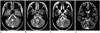
A 26-year-old female presented with recurrent generalized tonic-clonic seizures occurring approximately at two-month intervals in past three years. The seizures lasted 5 to 10 minutes with altered consciousness, which was brought under control with anti-convulsants. At admission, she was alert and was able to follow commands. There were no neurologic deficits at the initial neurologic examination, and electroencephalogram and laboratory findings were normal. She underwent MR imaging using 3 T system. On MR imaging, folia of the right cerebellar hemisphere showed an abnormal vertical orientation with smaller size of the right cerebellar hemisphere than that of the left side. In addition, the corticomedullary junction of the affected cerebellar hemisphere shows irregular and nodular configurations with indistinct margin (Fig. 1). Foliation of the left cerebellar hemisphere, formation of the vermis and configuration of the fourth ventricle were normal, as were the supratentrial structures, except for partial agenesis of corpus callosum (posterior body and splenium).
DISCUSSION
In previous reports, minor cerebellar dysplasias are often observed in the white matter and nodulus of the vermis in healthy newborns. These abnormalities are common features of human cerebellar development, and may persist during adult life (5, 6). However, major cortical cerebellar dysplasias have been reported in congenital muscular dystrophies and related disorders, or in intrauterine infection (1-3, 7). In such conditions, abnormal orientation of the cerebellar folia is secondary to the combined critical developmental abnormality of the cerebellum such as absence or hypoplasia of the vermis, or midline fusion abnormality (1-4, 7).
To our knowledge, there are only two reports that described isolated unilateral cerebellar cortical dysplasia showing vertically oriented folia and fissure, and the incidence is still unknown. Among total 9 cases of isolated unilateral cerebellar cortical dysplasia, 8 cases showed other associated malformations in the supratentorial structures (4, 5). In our case, the patient had a partial agenesis of corpus callosum involving posterior body and splenium. Demaerel et al. (3) reported a similar case, but the lesion was bilateral.
Understanding this abnormality requires knowledge of cerebellar cortical developments from embryological perspectives. It is well-known that cerebellar cortical neurons have dual origins. Up to about 10 weeks of gestation, the neural cells that forms the deep nuclei and the Purkinje layer of cerebellar cortex migrate radially outward from the germinal matrix. In contrast, at around 10 to 11 weeks, the neurons migrate tangentially over the cerebellar surface to form the granular layer (5, 8). The early signs of foliation, manifesting prior to the fissure formation, are locally increased in premigratory granular cells and indentation of the Purkinje cell layer. Up to the 40th week, the cerebellar lamellae are made up of four layers: the external granular layer, the molecular layer, the Purkinje cell layer, and the internal granular layer. During the first 6 to 8 months of extrauterine life, the external granular layer subsides progressively as its cells migrate inwards and the cerebellar lamellae assume their adult appearance within only three layers (5, 9). The external granular layer, Purkinje cell layer, and the overlaying meningeal cells may be involved in the mechanism of foliation (5, 10). The abnormal aggregation of external granule cells is preceded by an aberrant migration and misorientation of Purkinje cells in the hemispheres and a disturbed arrangement of glial fibers, suggesting that Purkinje cell settlement may be the key in cerebellar development (5, 6).
We report a rare case of isolated unilateral cerebellar cortical dysplasia with partial agenesis of corpus callosum with subtle changes on MR imaging. Our case suggests that awareness of this abnormality and its embryologic background can be helpful for recognition of this mild form of cerebellar abnormality on high-quality MR imaging in clinical practices.
 XML Download