

 PDF
PDF ePub
ePub Citation
 Print
Print


INTRODUCTION
Dysplasia epiphysealis hemimelica (DEH) or Trevor's disease is a rare non-hereditary developmental bone dysplasia characterized by an osteocartilaginous tumor arising from an epiphysis (1). The general prevalence has been reported be 1 in 1000000 and the etiology is unknown. Most patients are first seen between the ages of 2 and 14 years and three times more common in boys (2). DEH predominantly occurs in the lower limb with the talus, distal tibia, distal fibula, distal femur, proximal tibia and tarsal bones in the order of the frequency (3).
CASE REPORT
Case 1
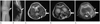
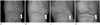
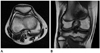
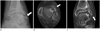
A 4-year-old boy presented to the orthopedics clinic, with incidentally found mass on the right knee with no symptoms. There was no history of trauma or previous joint swelling. The patient had normal gait and normal range of motion of the knee. Radiographs of the right knee showed multiple ossifications in the postero-medial aspect of the distal femoral epiphysis. The metaphyses of the femur and tibia were normal (Fig. 1A). Ultrasound and magnetic resonance imaging (MRI) revealed the presence of asymmetric epiphyesal cartilaginous overgrowth, which contained multiple ossifications (Fig. 1B-E). It was decided to observe the patient with no surgical intervention since no pain and normal range of movement of the knee.
Case 2
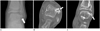
A 10-year-old boy presented to the orthopedic clinic, with ankle pain and mass on the anterolateral aspect of the left ankle, which was palpable for a month. There was a history of left ankle sprain about 8 months ago. Physical examination revealed a palpable hard mass (1 × 1 cm in size) of the anterior lateral aspect of the talus. Pain was induced on full dorsiflexion. Radiostagraph and computed tomography (CT) of the ankle showed excessive bony overgrowth from the anterolateral aspect of the talus with soft tissue swelling (Fig. 4). The abnormal over-growth portion of the talus was excised and for pathological examination which indicated dysplasia epiphysealis hemimelica. Two years later, the patient revisited with ankle pain. Radiograph and CT of the ankle revealed recurrence of the dysplasia epiphysealis hemimelica (Fig. 5). Surgical resection was repeated for the recurred mass with similar pathological results as before.
DISCUSSION
The clinical manifestations of Trevor's disease can be varied. The most common presenting complaints are from a painless deformity around a joint to a painful joint with mechanical symptoms (1). The typical radiographic finding is asymmetric epiphyseal cartilaginous overgrowth, containing multiple ossification centers. The patterns of the epiphyseal chondral calcification are variable namely stippled, irregular or dense. The epiphyseal calcified spots of the lesion are often multi-centric, which are gradually enlarged with mineralization and become confluent with the main epiphysis. CT is useful for the detection of small foci of early calcification or ossification within the cartilaginous mass and can identify cortical and medullary continuity between the DEH lesion and the adjacent bone. MRI is the technique of choice to identify the dimensions of the unossified cartilage mass, the extent of epiphyseal involvement and the status of the epiphysis (4).
Differential diagnoses include myositis ossificans, infection, tumoral calcinosis, synovial chondromatosis, loose bodies, vascular or parasitic calcification on radiography. Biopsy is not necessary; however, if imaging results are not conclusive, biopsy should be performed to exclude chondrosarcoma and osteosarcoma (5).
Management options for the treatment of Trever's disease include simple observation or surgical excision. By considering pain or deformity, appropriate treatment should be performed. As in our first case, asymptomatic DEH can be observed because there is no known risk of malignant transformation. Prognosis and symptoms depend on the site and size of the lesion and the degree of incongruity of the involved site. The recurrence rate of the deformity is reported to be high (6).
The two cases presented here were precisely diagnosed by CT or MRI prior to the management. Since the different symptoms and deformities of the each patient, the first was observed for eight years and the other was surgically managed. In particular, there is significant that first case was confirmed by radiologic image that DEH findings gradually became confluent with the main epiphysis during observation for eight years and finally made normal bone contour and density without deformity.
 XML Download