

 PDF
PDF ePub
ePub Citation
 Print
Print


INTRODUCTION
Acute compartment syndrome of the lower leg is a surgically emergent condition in which the pressure within the anatomically closed osseofascial compartment rises to reduce arterial and capillary circulation, finally leading to irreversible muscle and nerve damage. It is usually associated with major trauma such as fracture or crushing injury, and most commonly occurs in anterior or deep posterior compartments. The acute isolated lateral compartment syndrome is less typical and usually associated with minor trauma. However, there are few reports of idiopathic acute isolated lateral compartment syndrome, and there is no report of its radiologic findings (1-3). This study presents a magnetic resonance imaging of a case in a 48-year-old female affected with idiopathic acute isolated lateral compartment syndrome of the lower leg with ipsilateral peroneal nerve palsy as a complication, which shows localized enlargement of the peroneal muscles with peripheral convex bowing and change of their signal intensity with fluid signal along the adjacent fascial planes.
CASE REPORT
A 48-year-old female patient presented in the emergency room complaining of a gradually worsening pain of the left lower leg and dorsum of the left foot, which began two days prior. The patient denied any trauma of the lower leg or undue exertion. The patient also denied any past medical history, including neurologic or vascular diseases.
On physical examination, the patient demonstrated a mild swelling of the left lower leg compared with the opposite side. She complained of severe pain when the ankle was passively rotated. Passive dorsiflexion of the left ankle produced significant pain at the anterior aspect of the ipsilateral lower leg and anterolateral aspect of dorsum of the ipsilateral foot. The motor power of tibialis anterior and extensor hallucis longus muscles of left lower leg were 0/5 and 4/5, respectively (grade 0: no muscle movement; grade 4: movement against resistance, but less than normal; grade 5: normal strength). The sensation of the left lower leg was intact, and the arterial pulses of ipsilateral dorsalis pedis artery and posterior tibial artery were palpable. The laboratory evaluation showed white blood cell count of 10450 (mm3, poly 84%) and the erythrocyte sedimentation rate of 33 (normal range : < 20 mm/hr). The level of serum creatine kinase was also increased (56.9 ng/mL). The serum glucose level, platelet count, prothrombin time, and activated partial thromboplastin time were within the normal range.
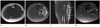
In the initial conventional radiographs of the left lower leg, there was no evidence of fracture or other bony abnormalities. The magnetic resonance (MR) imaging was taken for further evaluation. The patient was examined by using 1.5 T MRI scanner (MAGNETOM Avanto, Siemens Healthcare, München, Germany). The fat-saturated T2-weighted spin echo sequence had the parameters of repetition time 3800/echo time 80. The MR imaging revealed marked swelling of the peroneus longus and brevis muscles (Fig. 1A). Diffuse high signal intensity (SI) on T2-weighted image (T2WI) was noted throughout the proximal two-thirds of the peroneus longus and brevis muscles. Fluid layer was noted within the surrounding deep and superficial fascial planes. Focal increased T2-high signal intensity suspected as reactive edema was noted in the extensor digitorum longus and soleus muscles abutting the peroneus longus muscle (Fig. 1B, C). On axial gadolinium-enhanced T1-weighted image, heterogeneous enhancement was noted in the affected muscles of the lateral compartment (Fig. 1D).
Considering clinical and MR findings, a preliminary diagnosis was anterior and lateral compartment syndrome with peroneal nerve palsy and the patient was transferred to the operating room. The color of the peroneus longus and brevis muscles was brownish, and showed necrosis with the loss of activity of the muscles. The left common peroneal nerve was compressed between the swollen musculature of left lateral compartment and proximal fibula. The musculature of ipsilateral anterior compartment was normal, and there was no evidence of intramuscular hematoma or muscular rupture. Considering the clinical findings, MR findings, and intraoperative findings, the patient was given a diagnosis of left common peroneal nerve palsy due to acute isolated lateral compartment syndrome. The patient underwent the emergent decompressive fasciotomy of left lateral compartment and neurolysis of left common peroneal nerve. The skeletal muscle biopsy of lateral compartment was done, but the result was non-specific showing patchy degenerating muscle fibers. The microbiological culture of the musculature of the lateral compartment was also negative. In the ongoing follow-up in the orthopedic clinic after a month, the patient was pain free, and the motor power of the left lower leg had recovered.
DISCUSSION
Acute compartment syndrome is a life-threatening condition and can be explained by arteriovenous gradient theory and ischemia-reperfusion injury. When the intracompartment pressure is elevated, capillary blood flow is compromised. Edema of the soft tissue within the compartment further raises the intra-compartment pressure, which compromises venous and lymphatic drainage of the injured area and leads to decreased tissue perfusion. Untreated compartment syndrome mediated ischemia of the muscles and nerves leads to eventual irreversible damage and death of the tissues within the compartment. On the other hand, in the setting of the prolonged ischemia in the osteofascial compartment, reperfusion can worsen the preexisting cellular damage and cause compartment syndrome (1, 4). In this case, the patient had neither the history of trauma nor condition that may have caused the prolonged limb ischemia.
The compartments of the extremity are anatomically-closed spaces bounded by fascia and bone. In the lower leg, there are four compartments which are anterior, superficial posterior, deep posterior, and lateral compartments. The acute compartment syndrome of the lower leg usually occurs in the anterior and deep posterior compartments. The isolated lateral compartment syndrome of the lower leg has been considered to be a rare condition (1-3).
The acute compartment syndrome of the lower leg has been described most frequently in association with major trauma such as open or closed fractures and crushing injuries (2). Atraumatic acute compartment syndrome has been associated with activity, myositis, and medical pathologies. The most common etiology for the atraumatic compartment syndrome is strenuous activity that may result in chronic exertional compartment syndrome (1). Various other etiologies for the atraumatic compartment syndrome have been reported, including the association with diabetes mellitus as microangiopathic disease (1-5). The etiology of the isolated lateral compartment syndrome include minor trauma such as athletic-play without obvious injury or ankle inversion injury. Some cases have been reported to have occurred spontaneously as an acute exacerbation of the chronic exertional lateral compartment syndrome (2).
In this case, the patient presented with acute compartment syndrome which was isolated to the lateral compartment. She had no history of trauma or underlying medical disease. The results of the laboratory test were within normal range, which ruled out coagulopathy, infection, and diabetes mellitus. The histologic results were nonspecific with no definite evidence of microangiopathic disease. In view of the above, the authors could not identify a specific cause for the acute compartment syndrome in the patient.
The confirmation of diagnosis for compartment syndrome has been made by measurement of intracompartmental pressure, which is invasive and painful, and has certain risks such as bleeding or infection. The diagnosis of a compartment syndrome is primarily clinical and supplemented by measurement of intracompartmental pressure (1). In fact, the pain with passive stretching of the muscles and pain that is out of proportion to the associated condition are the most sensitive and reliable findings for compartment syndrome (1). If the diagnosis of a compartment syndrome is clinically apparent, measurement of intracompartmental pressure is not necessary and likely reserved for uncooperative patients or equivocal cases (6). In the present patient, she presented significant level of pain with passive stretching of the muscles, although she had history of neither trauma nor medical disease.
MR imaging has been used as a noninvasive method enabling examination of muscles. According to several studies, MR imaging is a promising technique for noninvasive diagnosis of compartment syndrome (7, 8). In acute compartment syndrome, MR imaging demonstrates diffuse hyperintensity on T2WI within the cross-sectional anatomic boundaries of affected compartment muscles (9). This hyperintensity on T2WI would appear to be the result of increased extracellular fluid in the affected muscle tissues by ischemic defects in the membrane permeability of capillaries (7). MR imaging also demonstrate swelling of affected compartment muscles with peripheral convex bowing, and hyperintense fluid or hemorrhage between the muscles in the fascial planes on T2WI (9). MR images of the current patient revealed marked swelling of the peroneus longus and brevis muscles within the whole length and lateral bulging of the affected muscles. Diffuse hyperintensity on T2WI was noted throughout the proximal two-thirds of the peroneus longus and brevis muscles. The fluid SI was noted within the surrounding deep and superficial fascial planes.
Diffuse hyperintensity of affected muscles on T2WI may be seen in various diseases and conditions (9). Among them, the differential diagnosis to be considered for the current patient is low grade muscle strain, nonspecific myositis, and muscle denervation edema. Low grade muscle strain may reveal diffuse edema, but usually along the musculotendinous junction. More severe muscle strains may contain fluid collection such as hematoma and interrupted muscle fibers and may show mass-like lesions. Nonspecific myositis includes infectious myositis and autoimmune inflammatory conditions such as polymyositis and dermatomyositis. These groups of myositis reveal nonspecific diffuse hyperintensity in the affected muscles on T2WI. As such, the correlation between MR images and clinical manifestation need to be considered to differentiate the diffuse hyperintensity of nonspecific myositis. Infectious myositis, especially bacterial myositis, may result from direct extension of infection in tissues adjacent to a muscle, such as osteomyelitis or subcutaneous abscess. Bacterial myositis frequently progress to the abscess formation. Autoimmune inflammatory conditions such as polymyositis and dermatomyositis are characterized by gradual onset of muscle weakness in the thighs and pelvic girdle that typically progresses to involve the upper extremities. Muscle denervation also causes edema diffusely throughout an involved muscle. However, the denervated muscle does not demonstrate change of signal intensity at MR imaging until 2-4 weeks after the denervation has occurred (9).
The treatment for compartment syndrome is emergent decompression of affected compartment by fasciotomy. If emergent decompression is delayed, irreversible muscle and nerve damages may occur within hours (2, 6).
We have reported the radiologic findings of the acute isolated lateral compartment syndrome of the lower leg without an obvious cause. In conclusion, although the measurement of the intracompartmental pressure is required for the definite diagnosis of acute compartment syndrome, such is not always necessary if the clinical and MR findings provide a reasonably high suspicion for the acute compartment syndrome (1, 6). The diagnosis of acute compartment syndrome should be considered in the following cases, even if the patient has no obvious cause: when the MR images demonstrate diffuse hyperintensity on T2WI within the cross sectional anatomic boundaries of the affected compartment muscles; the swelling of affected compartment muscles with peripheral convex bowing; and hyperintense fluid or hemorrhage between muscles in the fascial planes on T2WI.
 XML Download